|
International Journal of Bioelectromagnetism Vol. 6, No. 2, pp. 27-32, 2004. |
 |
 |
www.ijbem.org |
|
Spatiotemporal Dipole Imaging of Junichi Horia, Bin Heb
aDepartment of Biocybernetics, Niigata University, Niigata-shi, Niigata, Japan Correspondence: J. Hori, Department of Biocybernetics,
Niigata University, 8050 Ikarashi-2, Niigata 950-2181, Japan. Abstract.
Cortical dipole imaging technique, which attempts to estimate the cortical dipole
distribution from the scalp potentials, is one of the spatial enhancement techniques.
In this approach, an equivalent dipole source layer is used to model brain electrical
activity and has been shown to provide enhanced performance in imaging brain
electrical sources as compared with the smeared scalp-recorded EEG. We explore
suitable spatiotemporal filters for inverse estimation of an equivalent cortical
dipole-layer distribution from the scalp potentials for imaging of brain electric
sources. We have developed the parametric projection filter (PPF) based cortical
imaging technique, which allows estimating cortical dipole layer inverse solutions
in the presence of noise covariance. We have expanded the PPF to the time-varying
filter in order to handle the spatiotemporally varying nature of brain electrical
activity. Concretely, the noise covariance and the regularization parameter of the
PPF are supposed to be time-variant in order to eliminate the influence of the
background noise and eyes blink artifact. The present simulation results indicate
that the estimation error is reduced substantially by taking the spatio-temporal
properties of the noise into consideration, such as eyes blink artifacts and the
proposed time-variant PPF method provides enhanced performance in rejecting
time-varying noise.
Keywords: High-Resolution EEG; Inverse Problem; Spatiotemporal Inverse Filter; Equivalent Dipole Sources; Cortical Dipole Imaging; Parametric Projection Filter; Artifact Elimination
1. Introduction It is of importance to obtain spatio-temporal information regarding brain electrical activity from noninvasive electromagnetic measurements. Because of inherit high temporal resolution of electroencephalogram (EEG) measurements, high resolution EEG imaging, which aims at improving the spatial resolution of the EEG modalities, has received considerable attention in the past decades. Such EEG imaging modalities would facilitate noninvasive localization of foci of epileptic discharges in the brain, and the characterization of rapidly changing patterns of brain activation. A number of efforts have been made to achieve high resolution EEG imaging. Among them of interest is the spatial enhancement approach, which attempts to deconvolve the low-pass spatial filtering effect of volume conduction of the head (for review see [Dale and Sereno, 1993]). Cortical dipole layer imaging technique, which attempts to estimate the cortical dipole distribution from the scalp potentials, is one of the spatial enhancement techniques. In this approach, an equivalent dipole source layer is used to model brain electrical activity and has been shown to provide enhanced performance in imaging brain electrical sources as compared with the smeared scalp EEG [Hori and He, 2001; He et al., 2002; Wang and He, 1998]. The inverse problem of EEG is ill-posed and in general a regularization procedure is needed in order to obtain stable inverse solutions. We have previously developed the parametric projection filter (PPF) based cortical dipole layer imaging technique, which allows estimating cortical dipole layer inverse solutions in the presence of noise covariance [Hori and He, 2001; Hori and He, 2003]. Our previous results indicate that the results of the PPF provide better approximation to the original dipole layer distribution than that of traditional inverse techniques in the case of low correlation between signal and noise distributions. In the present study, we have expanded the PPF inverse spatial filter to the time-varying filter in order to handle the spatiotemporally varying nature of brain electrical activity. Concretely, the noise covariance and the regularization parameter of the PPF are supposed to be time-variant in order to eliminate the influence of the background noise and eyes blink artifact. 2. Methods 2.1 Spatiotemporal Dipole Layer Source Imaging The observation system of brain electrical activity on the scalp shall be defined by the following equation:
where fk is the equivalent source distribution of a dipole layer (DL), nk is the additive noise and gk is the scalp-recorded potentials. Subscript k indicates the time instant. A denotes the transfer matrix from the equivalent source to the scalp potentials. It is important to estimate the origins from the scalp-recorded EEG, and to image the sources that generate the observed EEG. The inverse process shall be defined by
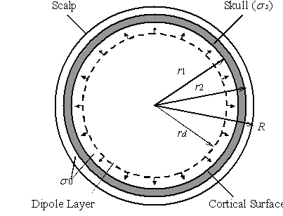
where Bk is the spatiotemporal restoration filter and f0k is the estimated source distribution of the DL. If the statistical information of the noise or signal are known or are estimated in accuracy, the restorative ability of the restoration filter Bk should be improved by using not only the transfer function but also the signal and noise information. In the present simulation study, the head volume conductor is approximated by the inhomogeneous three-concentric-spheres model [Wang and He, 1998]. This head model takes the variation in conductivity of different tissues, such as the scalp, the skull and the brain, into consideration. An equivalent DL is assumed within the brain sphere being concentric to the cortical surface. Radial current dipoles are uniformly distributed over the spherical DL to simulate brain electrical sources accounting for the scalp potentials. The electrical sources inside of the DL sphere are equivalently represented by the DL surrounding the sources, regardless of the number or the direction of the dipole sources [Hori and He, 2001; He et al., 2002; Wang and He, 1998]. The transfer function from the DL to the scalp potentials is obtained by considering the geometry of the model and physical relationship between the quantities involved. The strength of the DL is estimated from the noise-contaminated scalp potentials. 2.2 Time-Varying Parametric Projection Filter When the statistical information of noise is presented, the projection filter can be applied to the inverse problem. Suppose Qk the noise covariance, which can be derived from the expectation over the noise {n} ensemble, E[n n*]. n* is the transpose of n. The parametric projection filter (PPF) [Hori and He, 2001; Hori and He, 2003] is derived by
with γk a small positive number known as the regularization parameter. The PPF, using the free parameter, can improve the restorative ability from the projection filter, which provides the orthogonal projection of the original signal onto the range of the restoration filter that minimizes the expectation over the noise component in the restored signal. We have applied the time-invariant version of the PPF to the cortical dipole layer source imaging [Hori and He, 2001] and cortical potential imaging [Hori and He, 2003]. The time-variant PPF can also be applied to the spatio-temporal inverse problem described by (2) [Hori at al., 2004]. We have developed a criterion for determining the optimum parameter. One possibility is to use the following cost function:
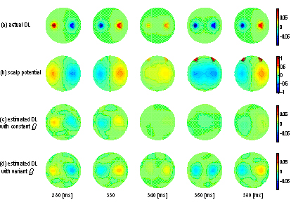
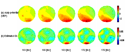
where f0k is the restored DL distribution using an initial value for γk, which should be relatively large to reduce the effect of additive noise on the coefficients. Furthermore, the recursive procedure that renewing the DL distribution f0k provides the optimum approximation of parameter γk. If there is no spontaneous artifact in the series of EEG measurements, the noise covariance Qk should be constant and it may be estimated from data that is known to be source free, such as pre-stimulus data in evoked potentials in a clinical situation [Sekihara and Scholz, 1995]. If there are some artifacts such as eyes blink, Qk should be adaptive to the spatial distribution of the artifacts in order to suppress them. The eyes blink covariance is substituted by the voluntary wink data. The eyes blink artifacts may be eliminated by using two types of noise covariance in the time-variant PPF according to the signal conditions with or without artifacts. The time-interval of eyes blink is estimated by the correlation coefficient between each scalp potential distribution and the eyes blink template measured by voluntary wink data in advance. 2.3 Simulation Protocol In the present simulation, two dipole sources were used to represent multiple localized brain electrical sources. The dipoles were oriented radially to the sphere with varying strengths. The strength of each dipole is changed from -1 to 1 with sinusoid in time. The frequencies of fluctuation in the dipole moments were set to 10 Hz and 30 Hz that assuming EEG alpha and gamma activities, respectively. In the present study, the source-conductor model [Wang and He, 1998; He, 1998; He, 1999] as shown in Fig. 1, was used. In this model, the radii of the brain, r1 , the skull, r2 , and the scalp, R, spheres were taken as 0.87, 0.92, and 1.0, respectively [Rush and Driscoll, 1969; Wang and He, 1998]. The normalized conductivity of the scalp and the brain was taken as σ0 = 1.0, and that of the skull as s= 0.0125. The potentials on the scalp surface, generated by current dipoles inside the brain, can be calculated by solving the forward problem from the dipole source to the scalp-surface potential [Kavanagh, et al., 1978]. The strength of the DL can be calculated by solving the forward problem from the assumed dipole source to the equivalent DL strength [He et al., 2002]. Two kinds of additive noise were considered. One is the time invariant noise such as a background noise that stochastic characteristics do not change in time. The time invariant background noise is expressed by Gaussian white noise (GWN). The noise level defined as the ratio between the norm of noise and the averaged norm of the simulated scalp potential over time was set to 0.1. The other is the time variant noise such as eyes blink artifacts. The movements of eyeballs generate electrical artifacts because the cornea sides of each eyeball are positively electrical-charged against the retina sides. The eyes blink artifacts appear as spike-like shape at upper parts of the eyes with the time duration of about 0.3 s. Since the amplitude of the eyes blink artifacts is at the order of 100 μV that is much greater than ordinary scalp potentials, the scalp measured potentials are degraded by the eyes blink artifacts. Figure 1. Schematic illustration of the head volume conductor-source model. The head is represented by an inhomogeneous concentric three-sphere volume conductor model with radii r1 , r2 , and R being 0.87, 0.92, and 1.0, respectively. The normalized conductivity of the scalp and the brain is taken as 1.0, and that of the skull as 0.0125. Dipoles are uniformly distributed over a sphere with the radius of rd. 2.4 Human Experiments Human visual evoked potential (VEP) experiment was carried out to examine the performance of the proposed restoration method. One healthy subject was studied in accordance with a protocol approved by the Institutional Review Board of the University of Illinois at Chicago. 96-channel VEP signals referenced to right earlobe were amplified with a gain of 500 and band-pass filtered from 1 Hz to 200 Hz, and were acquired at a sampling rate of 1 kHz. Half visual field pattern reversal check boards with reversal interval of 0.5 sec served as visual stimuli and 400 reversals were recorded to obtain averaged VEP signals. 3. Results A DL with 1280 radial dipoles at a radius of 0.8 was used [Wang and He, 1998]. Figure 2 (a) shows one example of the actual DL distribution of two radial dipoles at several time points. The dipole sources were located at the eccentricity of 0.7 with the angle of π/3. The strength of each dipole is changed with sinusoid in time (10 Hz and 30 Hz). The scalp potential was contaminated with two kinds of additive noise (Fig.2 (b)). One is the time invariant background noise expressed by Gaussian white noise. The other is the time variant noise of eyes blink artifacts, which appear as spike-like shape at upper parts of the eyes. As shown in Fig. 2(d), the DL distribution obtained by the time-variant PPF shows two areas of well-localized activity similar to the actual DL source distribution and the artifacts were eliminated. The relative error between the actual and estimated DL distributions was reduced by the time-variant PPF in every time instant. Especially, the relative error during the period with the artifact was dramatically reduced. The pattern reversal VEP data at the P100 were analyzed by the restoration filters of the PPF. Figure 3 shows an example of the normalized scalp potential map and the estimated cortical dipole layer maps in a healthy subject. As shown in Fig. 3 (a) in response to the left visual field stimuli, dominant positive potential components were elicited with a widespread distribution on the bilateral scalp. However, the estimated cortical DL distributions reveal a dominant in the right visual cortex as shown in Fig. 3 (b). Figure 2.Cortical DL imaging of two radial dipoles. (a) Actual DL distributions. (b) Scalp potentials contaminated with artifact and noise. (c) Estimated results with constant Qk and time-variant γk. (d) Estimated results with time variant Qk and k. Figure 3. Visual evoked potentials and the estimated DL distributions of a healthy subject around P100. (a) Scalp potential maps in response to the left visual field stimuli. (b) Estimated results by the time-variant PPF. 4. Discussion Research progress in the past decade has established the high-resolution EEG methodologies for imaging brain electrical activity. The cortical imaging approaches are virtually applicable to any kind of brain source distribution (both localized and distributed) [He, 1999]. This is due to the generalized nature of the equivalent surface source models behind the cortical imaging techniques. These techniques should be useful particularly for localizing and imaging cortical sources. Noise plays an important role in cortical DL imaging, as in any other ill-posed inverse problem. In the present study, we have investigated the performance of cortical DL imaging by considering noise covariance through the use of time-variant PPF. The present study demonstrates that enhanced performance can be obtained in cortical DL imaging by considering the noise covariance. The present results suggest that, the proposed method is effective in improving performance of cortical imaging, under the condition of low correlation between signal and noise. The present method would have similar restorative ability to the regularization procedures without considering the information of noise covariance, under the condition of high correlation between signal and noise. Therefore, using the present PPF approach will improve the performance of cortical potential imaging, considering the general cases that there is no high correlation between signal and noise. If we can obtain both signal covariance matrix and noise covariance matrix, the parametric Weiner filter (PWF) may be applied to the inverse problem [Dale and Sereno, 1993; Philips, et al, 1997]. There is no, to our knowledge, comprehensive investigation on cortical DL imaging in the metric of noise, in which non-white noise is considered. Actually, it is difficult to estimate the signal covariance exactly from the observed scalp potentials. Whenever the signal covariance is estimated, the PWF reconstructs the averaged signal over the time. We have confirmed the restorative ability of the PPF and the PWF in a separate study [Hori at al, 2002]. The simulation results obtained in that study suggest that, the PPF has better performance than other inverse filters under the condition of low correlation between signal and noise distributions. On the other hand, the PWF with incorporating signal information provides good results of equivalent dipole source imaging results compared to the PPF and Tikhonov regularization without signal covariance under the condition of high correlation between signal and noise distributions. As mentioned above, since the correlation between the eyes blink artifact and the brain electrical activities are low in most cases, we may use the PPF for eyes blink artifact suppression. If we can obtain the signal covariance in time course, the time-variant PWF would be also applicable to the equivalent cortical dipole layer imaging. In order to improve the resolution of restored dipole source imaging, we should choose the PPF and PWF according to the correlation between signal and noise distributions. The time variant PWF will be addressed in future investigation. 5. Conclusion We have developed a time-varying noise covariance incorporated inverse filter for cortical imaging, and showed its applicability in suppressing rapidly changing artifacts. The present simulation results suggest that the estimation error is reduced substantially by taking the spatio-temporal properties of the noise into consideration, such as eyes blink artifacts. Moreover, the VEP experimental study demonstrated that enhanced performance can be obtained in cortical imaging by considering the noise covariance. Further investigations on other applications of this new method should be addressed in the future. Acknowledgements This work was supported in part by Grant-In-Aid for Scientific Research from Japanese Society for the Promotion of Science No. 13480291, Grant for Promotion of Niigata University Research Projects, NIH 1RO1EB00178, and NSF BES-0411898. References
Dale AM, Sereno MI. Improved localization of cortical activity by combining EEG and MEG with MRI cortical surface reconstruction: a linear approach. Journal of Cognitive Neuroscience, 5: 162-176, 1993. He B. High resolution imaging of brain electrical sources. IEEE Engineering in Medicine & Biology, Sept./Oct., 123-129, 1999. He B. Brain Electric Source Imaging - Scalp Laplacian mapping and cortical imaging. Critical Review in BME 27: 149-188, 1999 . Hori J, He B. Equivalent dipole source imaging of brain electric activity by means of parametric projection filter. Annals on Biomedical Engineering, 29: 436-445, 2001. He B, Yao D, Lian J. High resolution EEG: on the cortical equivalent dipole layer imaging. Clinical Neurophysiology, 113: 227-235, 2002. Hori J, Lian J, He B. Comparison between parametric Weiner filter and parametric projection filter in cortical equivalent dipole layer imaging. In proceedings of 2nd Joint EMBS/BMES Conference, 2002, 929-930. Hori J,. He B. EEG cortical potential imaging of brain electrical activity by means of parametric projection filters. IEICE Transactions on Information & Systems, E86-D: 1909-1920, 2003. Hori J, Aiba M, He B. Spatio-temporal dipole source imaging of brain electrical activity by means of time-varying parametric projection filter. IEEE Transactions on Biomedical Engineering, 51: 768-777, 2004. Kavanagh RN, Darcey TM, Lehmann D, Fender DH. Evaluation of methods for three-dimensional localization of electrical sources in the human brain. IEEE Transactions on Biomedical Engineering, 25: 421-429, 1978. Philips JW, Leahy RM, Mosher JC, Timsari B. Imaging neural electrical activity from MEG and EEG. IEEE Transactions on Medical Imaging, 16: 338-348, 1997. Rush S, Driscoll DA. EEG electrode sensitivity - An application of reciprocity. IEEE Transactions on Biomedical Engineering, 16: 15-22, 1969. Sekihara K, Scholz, B. Average-intensity reconstruction and Weiner reconstruction of bioelectric current distribution based on its estimated covariance matrix. Transactions on Biomedical Engineering, 42: 149-157, 1995. Wang Y, He B. A computer simulation study of cortical imaging from scalp potentials. Transactions on Biomedical Engineering, 45: 724-735, 1998.
|