 |
International Journal of Bioelectromagnetism Vol. 5, No. 1, pp. 318-319, 2003. |
www.ijbem.org |
|
Model of Heart Shape Cyclic Variation
for Sergei Malchenko and Jüri Vedru University of Tartu, Tartu, Estonia Correspondence: Jüri Vedru, University of Tartu, Tartu, Estonia. E-mail: vedru@ut.ee, phone +372 7 380292 Abstract. A computer model
has been developed that provides a realistic description of the deformation
of the human heart surface shape during a single cardiac cycle. The model
is proposed for bioimpedance measurement simulations.
Keywords: Epicardium; Spline Surface; Foucault Cardiography; Electrical Bioimpedance 1. Introduction Foucault Cardiography (FCG) [Humal et al., 1996] is an electrical bioimpedance method being developed at the University of Tartu for tracking the heart mechanical activity. It is based on the influence of the variations of heart shape and position onto the electromagnetic power absorption in the thorax. To take more precise notion about the advantages and disadvantages of the FCG, a computer model describing the variation of the human heart shape is necessary. 2. Heart Surface Model Composition The model of heart surface variation was composed on the basis of a set of real time MRI images, applying also physiological knowledge. The initial MRI data for the work model was obtained from [Hoffman et al.]. The initial data comprises a movie of the heart beating: 160 images data set taken at 10 time points for 16 sections of the heart. 2.1. Main Principles In the process of reconstruction the following principles have been considered. We had to reconstruct of the heart surface (epicardium) and its variation during a heart cycle without taking into account the inner structure of the heart. Specific formations of the heart surface near the juncture of the heart with the blood vessels were not described. Anatomic and functional connections of pericardium with adjacent organs also have not been taken into account. Main physiological facts (e.g., correlation between the duration of systole and diastole, proportions of the axial and radial dimensions of the heart, known forms of volume curves) were taken into account. 2.2. Heart Surface Reconstruction and Adjustment We have got the collection of images for every time moment: ten time moments and sixteen images for each time moment. For each image from this collection the corresponding contour of the heart was found. Using the program package MATLAB on each image, appropriate contours of the heart were marked, obtained and represented in the Cartesian coordinates. By means of a spline function a closed and continuous surface for each single time point was obtained. Fig. 1 represents heart surface construction for a single time moment.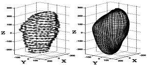 Figure 1. Composing of heart shape using spline surface. Left collection of contours, right obtained heart surface.
Using the described above approach the heart shapes for all ten time moments were obtained. Counting on the dependence of the epicardium shape on time, the time coordinate was introduced. We have adjusted the model and introduced additional time moments in it, for which additional shapes also were produced. The objective was to fit the time course to the facts known from physiology. The proportions of the whole heart and distances between heart sections were also adjusted. 3. Results For description of the heart surface, a spline function r = F (j, q, t), in spherical coordinates and time, was chosen [Malchenko S., 2002]. By the changing of the smoothing degree we have also an opportunity to receive different degrees of approximation of the form of pericardium. By the means of spline smoothing, the closed and continuous surface for all time points was obtained.
 Figure 2. Total heart volume (VT), total ventricular volume (VV) and total atrial volume (VA) variation (for convenience, shifted close to each other).
The model was used as the tool for calculation of cycle curves of ventricular, atrial and total heart volumes. Fig. 2 represents total heart volume VT, total ventricular volume VV, and total atrial volume VA variations. The movement of atrioventricular septum was taken into account at volume calculations. The model was also used as a tool for studying of small variations of the heart surface. The regions with maximum motions are located on the surface of ventricles; smaller motion can be found on the heart surface above the septum between the left and right ventricles. The motion in the in atrial regions is less and not so strongly marked. 4. Discussion We have developed an empirical model, where the modeled heart surface is closed and smooth and gives a possibility to track the variations of the heart surface. Generally, it looks well and is reliable. Basic features of the variations of the heart shape are detected. Total heart, ventricular and atria volume variation are in good correspondence with the facts, known from physiology. A particularly important phenomenon for FCG studies is plausible imitation of small variations of the heart shape. The latter determine the generation of Foucault cardiogram. That is why we have attached much importance to it. The obtained model was used as a tool for deriving a collection of cuts of the exterior surface of the heart which where used for FCG studies [Skaburskas et al., 2003]. But problems still remained. The model is a particular case and cannot be easily adjusted for some other individuality. The time course of the heart motion during the contraction cycle cannot be modulated. The model omits inner structure of the heart and connections to the great blood vessels. Motion of the heart in the thorax due to breathing is not described. Small, uncorrelated heart surface oscillations that are located near the base and the apex of the heart still remained. For further investigations more complete model is necessary. We have to introduce the simulation of the heart motion in the thorax due to breathing and add a description of the variation of the ventricles and atria. Also, by introduction of additional adjustable parameters it ought to be possible to fit existing heart shape to a desired form. Acknowledgements The authors thank Dr. Eric A. Hoffman, Ph.D., Professor of Radiology and Biomedical Engineering, Department of Radiology, University of Iowa College of Medicine, for kind permission to use the CT data; and the Estonian Science Foundation for supporting this work by its grant No 3914. References Humal L-H, Vedru J. Physiological measurement based on Foucault principle: set-up of the problem. Medical & Biological Engineering & Computing, 34, Suppl.1, Part 2: 183-184, 1996. Hoffman EA et al. Welcome to Physiological Imaging (website). Division of Physiologic Imaging, Universityof Iowa College of Medicine. http://everest.radiology.uiowa.edu Malchenko S. Model of human heart shape variation during cardiac cycle. MATLAB File Exchange (website). MathWorks. http://www.mathworks.com/matlabcentral/fileexchange, 2002. Skaburskas K, Vedru J. Origination of Foucault cardiogram by impedance redistribution: Transfer coefficient approach. In this volume.
© International Society for Bioelectromagnetism
|

