 |
International Journal of Bioelectromagnetism Vol. 5, No. 1, pp. 314-315, 2003. |
www.ijbem.org |
|
Methods for Using Ultrasound to Generate a Heart Surface for Electrocardiographic Inverse Problems Jason W. Trobaughab and
R. Martin Arthura aElectronic Signals and Systems Research Laboratory,
School of Engineering and Applied Science; Correspondence: RM Arthur, Department of Electrical
Engineering, Washington University in St. Louis, Campus Box 1127, Abstract. Errors of electrocardiographic
inverse solutions are especially sensitive to the accuracy with which a heart
model is constructed and placed within the torso. In an effort to reduce those
errors, we have improved our previous methods for generating individualized
heart models from ultrasound images. An ultrasound probe is tracked using
an Immersion digitizer, allowing measurement of the orientation and position
of each image. Images are tracked relative to the torso, which is also measured
using the digitizer. The probe is calibrated by imaging a phantom consisting
of multiple N-fiducials and computing a transformation between ultrasound
coordinates and Immersion measurements. Using these methods, we have constructed
a heart surface model from apical and parasternal images acquired at multiple
rotations and tilts with a Terason 2000 imaging system. Accuracy of the methods
is limited by multiple factors, including patient movement, tracking error,
and image analysis. We plan to investigate these limitations and to extend
these methods to model dynamic, time-varying heart shape and location. Such
studies are critical for the development of individualized anatomies, which
are crucial for continued advancement of inverse methods.
Keywords: Inverse Electrocardiography; Ultrasound; Calibration; Individualized Anatomy 1. Introduction 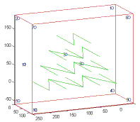 Figure 1. Sketch of calibration phantom consisting of four rows of N-fiducials and 11 registration divots (numbered circles). Divots enable mapping the phantom and Immersion coordinates, and the N-fiducials enable calibration of the ultrasound probe. Errors in torso and heart geometry are known to affect electrocardiographic inverse solutions. The extent of these effects is still a subject of research, however, as are the limitations of current methods for routinely obtaining accurate representation of anatomical structures in a clinical setting. We believe that subject-specific models would contribute significantly to the reduction of errors in inverse solutions. Towards that objective, we have used ultrasound for the construction of heart models [1]. Our current effort is aimed at improving previous methods and determining the limits of accuracy achievable with existing techniques. 2. Material and Methods To capture 3D heart geometry we used a TerasonTM 2000 ultrasound imaging system, coupled with an Immersion MicroScribe-3DL digitizer. The inexpensive imaging system consists of a laptop computer with imaging software, a 64-element, 2.5 MHz probe (4V2) and a beamforming module. The 3DL is a mechanical, articulated arm that reports 3D position and orientation of a stylus tip. For calibration of the ultrasound probe, we constructed a plexiglass phantom consisting of multiple N-fiducials as shown in Figure 1. To immobilize the subject, we use a Vac-LokTM, a cushion filled with polystyrene beads that forms rigidly to the patient when a vacuum is applied. Spatial positioning of multiple images requires measurement of the relative orientation and position of each image plane. We have implemented techniques developed initially for the field of image-guided treatments, e.g., [2]. The objective for each image plane is a mapping between the ultrasound image pixels and a reference 3D coordinate system such as that of the digitizer. A calibration step involves finding this rigid mapping, a rotation and translation, for some initial measurement of the stylus orientation and position. Subsequent digitizer measurements define the mapping for each new image. 3. Results
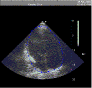 Figure 2. Typical apical four-chamber ultrasound image with points on the heart surface and cubic spline contour generated from the points.
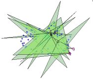 Figure 3. Relative geometry of multiple image planes and surface points used to construct the heart surface. Our cardiac probe was calibrated as described above, and images of a normal adult subject were obtained in multiple views. A typical apical four-chamber view is shown in Figure 2, with points overlaid that we identified as part of the heart surface. Analysis was performed with a side-by-side comparison of the static image shown here and a loop of images showing the cardiac cycle. This image was chosen at end-diastole as judged by visual inspection of motion in the image loop. For this subject, ten images were acquired, consisting of apical two- and four-chamber images, and parasternal long- and short-axis images at multiple view angles in an attempt to maximize coverage of the heart surface. The image planes are shown in Figure 3, in roughly the same orientation as the heart and torso surface shown in Figure 4. The heart surface points identified on each image are also plotted. The convex hull of the heart surface points shown in Figure 3 was generated to form a closed, triangulated surface. That surface is shown in Figure 4 inside a semi-transparent model of the subjects torso. Relative alignment of the two surfaces was known in this case based on identification of homologous points in the torso model and on the subject during data collection. In general, all torso and heart points would be acquired relative to the same reference coordinates.
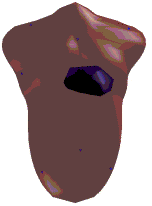 Figure 4. Individualized heart and torso surfaces. Relative alignment is based on anatomic fiducials measured during acquisition of each surface.
4. Discussion Our preliminary results show feasibility of these methods and potential for modeling heart and torso geometry for inverse electrocardiography in a clinical environment. Ultimately, these methods should produce limits on the accuracy achievable with available technology. Rigorous quantitative analysis must be done to thoroughly assess effects on inverse solutions. This analysis is made difficult by the lack of a gold standard and by the following factors: limited imaging windows, calibration and tracking errors, motion of the torso and heart associated with breathing, patient immobilization, analysis of cardiac images, and methods for generation of a surface from available image data. The studies are critical for the development of individualized anatomies for inverse electrocardiography, which are crucial for continued advancement of inverse methods. Acknowledgements This work was supported in part by National Institute of Health grant R01-50295 and by the Wilkinson Trust at Washington University. References [1] Arthur, R. M., Beetner, D. G., Ambos, H. D., Cain, M. E. Improved Estimation of Pericardial Potentials From Body-Surface Maps Using Individualized Torso Models, J. of Electrocardiology, 31(Suppl): 106-113, 1999. [2] Pagoulatos, N., Haynor, D.R. Kim, Y. A fast calibration method for 3-D tracking of ultrasound images using a spatial localizer. Ultrasound in Medicine and Biology, 27(9): 1219-1229, 2001.
© International Society for Bioelectromagnetism
|

