 |
International Journal of Bioelectromagnetism Vol. 5, No. 1, pp. 282-283, 2003. |
www.ijbem.org |
|
ST-T Isointegral Patterns in the
M Sobieszczanskaa,
J Jagielskia, L Rusieckia, W Kustrzyckib, and
J Oczkob aDepartment of Pathophysiology, Wroclaw Medical
University, Wroclaw, Poland Correspondence: M Sobieszczańska, Department of Pathophysiology, Wroclaw Medical University, Marcinkowskiego 1, 50-368 Wroclaw, Poland. E-mail: msob@patfiz.am.wroc.pl, phone +48 71 784 00 60, fax +48 71 784 00 61 Abstract. A goal of the study was
to investigate a possible relevance between the qualitative and quantitative
features of ST-T isointegral surface maps and the significant stenosis of
the single coronary artery in the patients with symptomatic CAD and without
specific ST-T abnormalities on the conventional 12-lead ECG. It was found
that the ST-T isointegrals minimum values were significantly deeper only in
the patients with LAD stenosis as compared with the controls. In the most
of the examined patients from the groups LAD, RCA and Cx, ST-T maps revealed
the specific distribution patterns. Thus, BSPM could be considered as a useful
supportive method in diagnosing myocardial ischemia not detectable with the
standard ECG.
Keywords: Single-Vessel CAD; Coronary Angiography; BSPM; ST-T Isointegral Maps 1. Introduction The patients with coronary artery disease (CAD) who do not reveal any discernible abnormalities on the standard 12-lead resting ECG constitute as much as even a half of the all CAD cases [2]. That group is still a subject of special interest regarding a search of diagnostic tools of the higher sensitivity. Body surface potential mapping (BSPM) was found to be a method characterized by a unique emphasis on spatial features of cardiac field, likewise by a documented, unequivocally better sensitivity to regional cardiac events without diminution of specificity [3], as compared with conventional ECG. We have attempted to investigate a correlation between the myocardial ischemic area, as reflected in the resting ST-T isointegral surface maps, and the stenosis confined to the single coronary artery in the patients with symptomatic CAD and with no concomitant changes on the conventional ECG recordings. 2. Material and Methods 2.1. Study Population A group of 84 patients with symptomatic coronary artery disease, after giving the informed consent, was enrolled to the study. There were 63 males and 21 females, whose age ranged from 39 to 74 years (mean: 56,48 (±11,08). The all patients reported a history of the typical angina pain episodes. The exclusion criteria were: previous Q-wave myocardial infarction, intraventricular conduction disturbances and ventricular hypertrophy. None of the examined patients revealed the specific alterations of ST-T on the 12-lead standard ECG recorded at rest. Treadmill exercise tests appeared to be positive in 63 of them, negative in 8, and 13 patients did not undergo exercise testing. Echocardiography examinations in the all cases showed no akinesis or severe dyskinesis. The all patients were subjected to coronary angiography procedure. According to the location of the critical stenosis (>70% luminal narrowing) isolated to a single coronary artery, the patients were divided into the three groups: LAD, RCA and Cx. ST-T isointegral surface maps obtained from the CAD patients were referred to the matched control group consisting of the 30 subjects with no evident cardiac involvement. 2.2. Method A procedure of BSPM was performed with a use of the HPM-7100 Fukuda Denshi system equipped with 87 recording electrodes covering the anterior and posterior thoracic surfaces. ECG signals were sampled simultaneously with the Wilsons central terminal as reference, at the rate of 1000 Hz, and averaged for ten cardiac cycles. Then the isointegral maps of ST-T were constructed by calculating for each lead the algebraic sum of the all instantaneous potentials throughout the interval ST-T, from J point to T wave offset instants, assigned from the edited Frank X, Y, and Z leads. ST-T maps depicted the thorax as a cylinder unrolled from the right axillary line with the front and the back framed by the A-I and J-M electrode rows, respectively. The patients with angiographically documented one-vessel CAD underwent the BSPM recordings at the rest and pain-free state. The analysis concerned the qualitative and quantitative features of the ST-T isointegrals maps obtained in the three CAD subgroups in relation to the normal ST-T pattern. 3. Results Distribution of the ST-T isointegral maps in the controls showed a smooth bipolar pattern, with a positive potential covering the precordial region, and a negativity located over the right chest and the entire back. A quantitative criterion considered was the group-mean value of the ST-T isointegral minimum extremes, which for the control group was established at -23,5 (±8,7) μVs. In the LAD group, comprising 24 patients, the mean value of minimum extremes was significantly deeper, i.e., -34,3 (±11,8) μVs, as compared with the controls. As to the potential localization, 18 patients of this group (75%) demonstrated the abnormal surface distribution, with a prominent negativity, replacing a normal positivity, appearing over the anterosuperior torso. This ST-T isointegral distribution pattern could be named anterior type. Regarding the RCA group, constituted by 42 patients, the mean isointegral minimum value was -29,7 μVs (±17,1). ST-T isointegral distribution in 35 of these patients (83%) differed considerably from the normal pattern, corresponding to the inferoposterior type, featured by the negative potential extended through the whole back and over the left and inferior portion of the thorax. The group with the isolated stenosis of Cx artery (18 patients) showed the mean minimum value of -18,2 μVs (±10,2). ST-T isointegral maps in 13 of these patients (77%) were found to have a distinct appearance from the normal maps, rendering the lateral type distribution, showing the negative areas located prominently over the left side of the thorax. 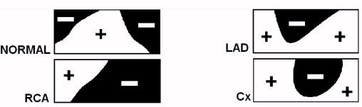
4. Discussion Application of BSPM for improving CAD diagnostics has been for years a subject of the multidirectional researches performed under different clinical and methodological conditions. The outcomes, as yet, remain controversial. ST-T configuration on ECG depends on the interrelation between subendocardial and subepicardial repolarization patterns. Coronary artery stenosis produce an insufficient perfusion of the subendocardial myocytes and accompanying ischemic fluctuations of their monophasic action potential, which results in ST-T changes [1]. Obviously, more evident ST-T maps abnormalities, including those indicating a site of ischemic region, are provoked by exercise testing [4, 5]. However, some authors reported also a usefulness of integral maps recorded at rest [2, 3]. It is in accordance with our findings suggesting that ST-T integral mapping could be of some value for identifying ischemia location in patients with single-vessel CAD. References 1. Flowers N.C., Horan L. Body surface potential mapping. In Cardiac Electrophysiology. From Cell to Bedside. Zipes D.P., Jalife J., Editors., 3rd ed., W.B. Sanders, Philadelphia, 2000, 737-746. 2. Green L.S., Lux R.L., Haws C.W. Detection and localization of coronary artery disease with body surface mapping in patients with normal electrocardiograms. Circulation, 76(6): 1290-1297, 1987. 3. Green L.S., Abildskov J.A. Clinical Application of Body Surface Potential Mapping. Clin. Cardiol., 18: 245-249, 1995. 4. Montague T.J. Exercise Body Surface Potential Mapping in Single and Multiple Coronary Artery Disease. Chest, 97(6): 1333-1342, 1990. 5. Nakajima T. et al. ST-T isointegral analysis of exercise stress body surface mapping for identifying ischemic areas in patients with angina pectoris. Am Heart J., 115(5): 1013-1021, 1988.
© International Society for Bioelectromagnetism
|

