 |
International Journal of Bioelectromagnetism Vol. 5, No. 1, pp. 279-281, 2003. |
www.ijbem.org |
|
Application of Wavelet Transform for
Analysis of Hisa Shimojimaa,
Takeshi Tsutsumia, Fumiko Yanagisawaa, Masae Komukaia,
Naoko Zendaa, aDivision of Cardiology, Showa University Fujigaoaka
Hospital, Yokohama 227-8501 Japan Abstract. This study was performed
to evaluate the frequency power spectrum during QRS in intraventricular conduction
abnormalities (IVCA). Electrocardiograms were recorded from normal volunteers
(n=12) and in IVCA (n=6) with or without myocardial infarction (MI). The signals
were inputted into Window based PC at sampling rate of 10kz as binary files.
To assess the frequency power of QRS, wavelet transform (WT) was utilized.
The time dependent changes in the frequency powers were extracted by use of
Morlet wavelet of 40 scales corresponding to frequency bands from 10 to 400Hz.
The time integral of the frequency power over the QRS and number of its peaks
during the time course (Sigma peak) were calculated. The results showed reduced
power spectrum at the frequency power (100Hz or less) was reduced in IVCA,
compared with normal cases. In IVCA with MI, Sigma peaks were increased at
high frequency range (150Hz or more). In conclusion WT is beneficial for the
analysis of signals within the QRS. Change in the power spectrum may show
the characteristics of cardiac excitation fronts.
Keywords: Wavelet Transform; Frequency Analysis; High Frequency Component; Myocardial Infarction; Intraventricular Conduction Abnormalities 1. Introduction It has been pointed out by several authors (Flowers, 1969; Maehara, 1999; Zenda 2000) that the scattered myocardial damages such as focal necrosis may be related to the production of high frequency component, but the clinical significance of these findings has not been established. Compared with widely used FFT algorithm, the wavelet transform (WT) is based on special functions with time-widths adapted to each frequency and is suitable for transient signal detection within the QRS (Morlet, 1993; Figliola, 1997), which can be applied for exploring the deformation of cardiac excitation wave front. It has been known that the progress of normal activation of the ventricular wall is nearly radial and that of intraventricular conduction abnormalities (IVCA) is more tangential. Different findings are expected in the power spectrum of these conditions, as well as in cases with conduction barriers such as myocardial infarction (MI). The present study was performed to evaluate the alteration of the frequency power during QRS in IVCA with or without MI by the new method using WT. 2. Material and Methods Standard 12 leads electrocardiogram (ECG) were recorded at sampling rate of 10 kHz from normal volunteers (n=12) and the patients with IVCA (n=6), and with IVCA plus MI. These signals were inputted into Window based PC as binary files. To assess the frequency power limited to QRS, WT was utilized for the assessment of signals within the QRS interval. The PC software was newly developed for this purpose (BIOMAS ver 1.0, Elmec Co., Inc., Tokyo, Japan). The Morlet wavelet was used as a mother wavelet. The time course of the signal power was obtained from 40 scales corresponding to frequency bands from 10-400Hz. In addition, the time integral of the power spectrum corresponding to each frequency was calculated over 100msec period from the onset of the QRS complex. These calculation steps were repeated on 10 consecutive beats and the results were averaged and compared in cases with and without MI. 3. Results 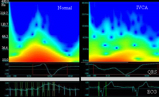 Figure 1. An representative cases of time-frequency relationships by use of wavelet transform. the vertical axis indicates the frequency, and the transverse one the time from the onset of QRS, surrounded by red frames. The color and its brightness show the magneitude of frequency power which is greater in red and smaller in green.
4. Discussion The results of this study indicate that the frequency analysis with WT is a beneficial tool for elucidating the frequency profiles of QRS complex. The reduction of the low frequency power in IVCA may be due to more tangential activation pattern of the ventricular wall. The increased power and number of peaks in high frequency range (150-250Hz) were observed in IVCA with MI, which may reflect the transformation of the excitation front passing throughout MI lesions. It is suggested that the methods provides a new approach to reveal the myocardial damage concealed under IVCA. 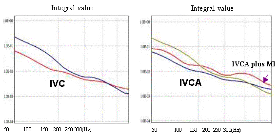 Figure 2. Time integral values of power spectrum against the every frequency band. The vertical axes show the integral values of frequency power calculated from each frequency band, and transverse axes the frequency, N: normal, IVCA: intraventricular conduction abnormalities, MI: myocardial infarction. References Flowers NC, Horan LG, Thomas JR, Tolleson WJ: The anatomical basis for high-frequency components in the electrocraiogram. Circulation XXXIX:531-539, 1969. Figliola A. and Serrano E: Analysis of physiological time series using wavelet transforms. IEEE Engineering in Medicine and Biology. May/Jun:74-79,1997. Langner PH, Geselowitz DB, Mansure FT: High-frequency components in the electrocardiogram of normal subjects and of patients with coronary heart disease. Am Heart J. 62:746-755, 1961. Morlet D, Peyrin F, Desseigne P, Rubel P: Wavelet analysis of high-resolution signal-averaged ECGs in postinfarction patients. J Electrocardiol 26:311-320, 1993. Maehara K, Kokubun T, Awano N, Taira K, Ono M, Furukawa T, Shimizu Y, Maruyama Y: Detection of abnormal high-frequency components in the QRS complex by the wavelet transform in patients with idiopathic dilated cardiomyopathy. Jpn Circ J 63:25-32, 1999. Zenda N, Tsutsumi T, Sato M, Takeyama Y, Harumi K, Wei D: Computer simulation of notches on initial part of QRS complex in patients with anterior myocardial infarction, in 2000 Electrocardiology. Ambroggi LD, Editor. Casa Editrice Scientifica Internazionale,Roma,2001,117-120.
© International Society for Bioelectromagnetism
|

