|
International Journal of Bioelectromagnetism Vol. 5, No. 1, p. 177, 2003. |
www.ijbem.org |
|
Investigating Heterogeneity
in Human Heart with Gunnar Seemann, Frank B. Sachse, Daniel L. Weiß, and Olaf Dössel Institute of Biomedical Engineering, Universität Karlsruhe (TH), Germany Correspondence: G Seemann, Institute of Biomedical
Engineering, Universität Karlsruhe (TH), Kaiserstr. 12, 76131 Karlsruhe,
Germany. Abstract. The electrophysiological
properties of cardiac tissue can be described with simulation environments
based on reaction-diffusion models. In this environment the reaction
term consists of ionic cell models and the diffusion term of a bidomain
approach. The underlying anatomical structure is a realistic representation
of human left ventricular tissue. As result transmural electrocardiograms
of the left ventricle are presented which show typical characteristics
of depolarization and repolarization in heterogeneous tissue. These
results are compared with those of homogeneous tissue. The presented
model will be used in future to simulate individual body surface
potential maps.
Keywords: Modeling; Simulation; Electrophysiology; Heterogeneity; Ionic Models; Bidomain Model; Human Ventricle 1. Introduction Precise knowledge of the physiologic and pathologic properties of the heart leads to a better understanding of heart function and therefore enables improvements in diagnosis and therapy. To enhance this knowledge computer simulations of the electro-mechanical properties are used. A complex model describing the electrophysiology of ventricular tissue is presented in this paper. This model is used to examine the effects of regional heterogeneity in the ventricular wall due to transmural changes of ion channel characteristics on transmural electrocardiograms (ECG). 2. Material and Methods The simulation environment is presented in this section and consists of three major components: cellular electrophysiology, excitation propagation and anatomical structure. For a more precise description, see [Seemann et al., 2002]. Cellular Electrophysiology: The electrophysiological behavior of a single cell is modeled with a modified Noble-Varghese-Kohl-Noble model, which was generated with measurements of ventricular cells of a guinea pig [Noble et al, 1998]. The model describes the electrical properties with a set of coupled, nonlinear partial differential equations of first order. These equations reconstruct intra- and extracellular ion concentrations, ion flows through the cell membrane, state variables and time dependencies of the ion channels, and the transmembrane voltage.
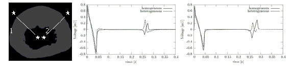
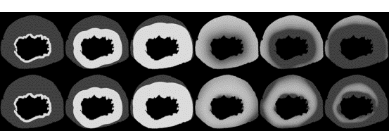 Figure 1. Gray-coded transmembrane voltage in a slice of the left ventricle during a heart cycle in a homogeneous (top row) and a heterogeneous (bottom row) model. Each first three pictures show the depolarization, the others the repolarization. Excitation propagation: Electrical coupling of cells by calculating the intercellular current flow reproduces the myocardium as a net of cells. Therefore, the bidomain diffusion model is used to represent the anisotropic current flow through gap junctions and the extracellular space [Henriquez et al, 1996]. The current flow is calculated with aid of Poisson´s equation for stationary electrical fields in two domains with the finite difference method. Anatomical structure: The realistic anatomical model bases on photographic images of the Visible Female Project [Sachse et al., 2000]. The images were transformed into a three-dimensional data set using techniques of digital image processing. The model includes via a rule-based method orientation of muscle fibers, which is needed to incorporate anisotropic electrical properties. In this work a slice of 3 x 257 x 216 voxels with a length of 0.33 mm is applied. Regional Heterogeneity: Transmural changes of ion channel characteristics of the currents Ito, IKs, IK1, and INaCa are mainly responsible for the regional heterogeneity of ventricular myocardium in human. These changes are assumed to be continuous from endocardium to epicardium. The heterogeneity is included with respect to measured data [Antzelevitch et al, 1999]. 3. Results Electrophysiological properties were calculated with a homogeneous and a heterogeneous model on a slice of the left ventricle. Depolarization and repolarization phase are shown in a transmural view in Fig. 1. No significant difference is visible during depolarization of the two models. The repolarization differs due to heterogeneous characteristics. While the end of repolarization vanishes in the homogeneous model at epicardium, in the heterogeneous it vanishes in midmyocardium. To further investigate these differences, Fig. 2 shows two transmural ECGs of each model. The ECGs of the homogeneous model have in both cases a biphasic T-wave. The heterogeneous model delivers a positive, nearly monophasic slope.
4. Discussion and Conclusions The presented heterogeneous model is suitable to simulate precisely the electrophysiology of ventricular tissue. The behavior of repolarization is investigated with simulated transmural ECGs. These ECGs correspond to measurements [Antzelevitch et al, 1999] illustrating a phenomenon, which is seen in specific leads of physiologic body ECGs, i.e. a monophasic positive T-wave. The model will be extended to simulate electro-mechanics of the complete heart. The simulations within this model improve the comprehension of pathologies and their influence on the cardiovascular system. Individual body surface potential maps will be simulated, to enable diagnostic advancements. References Antzelevitch C, Yan G, Shimizu W, Burashnikov A. Electrical heterogeneity, the ECG, and cardiac arrhythmias. Cardiac Electrophysiology. From Cell to Bedside. Zipes DP, Jalife J, Editors. W.B. Saunders Company, Philadelphia, 1999, 222238 Henriquez CS, Muzikant AL, Smoak CK. Anisotropy, fiber curvature, and bath loading effects on activation in thin and thick cardiac tissue preparations: simulations in a three-dimensional bidomain model. Journal of Cardiovascular Electrophysiology, 7, 424444, 1996 Noble D, Varghese A, Kohl P, Noble P. Improved guinea-pig venticular cell model incorporating a diadic space, IKr and IKs, and length- and tension-dependend processes. Canadian Journal of Cardiology, 14, 123134. 1998 Sachse FB, Werner CD, Stenroos MH, Schulte R, Zerfass P, Dössel O. Modeling the anatomy of the human heart using the cyrosection images of the visible female data set. In proceedings of the 3rd Users Conference of the National Library of Medicine's Visible Human Project. 2000 Seemann G, Sachse FB, Dössel O. Excitation Propagation and Force Development in the left Ventricle of the Visible Female Data Set. Biomedizinische Technik, 47 (1/1); 221224, 2002
© International Society for Bioelectromagnetism
|