|
Computer Aided Medical Image Diagnosis Enabling Electrophysiological Simulation Studies
Tomi
Heinonenab, Prasun
Dastidarc
aRagnar
Granit Institute, Tampere University of Technology, Tampere, Finland
bNokia Corporation, NVO, Tampere, Finland
cTampere University Hospital, Department of Diagnostic Radiology, Tampere, Finland
Correspondence: T Heinonen, Ragnar Granit Institute, Tampere University of Technology
P.O. Box 692, FIN-33101 Tampere, Finland.
E-mail: tomi.heinonen@nokia.com, phone +358 50 3738 611, fax +358 3 247 4013
 Abstract. Due to increasing number of PACS
installations and efficient computing environments the popularity of
computer-aided diagnosis is increasing rapidly. There are numerous applications
available for evaluating different disease signs. Many PACS manufacturers
develop multipurpose PACS workstation software enabling interactive image
displaying and analysis tools. In addition to obtaining useful
quantitative information for the analyzed images, the resulting data can
be applied in electrophysiological studies, i.e., segmented anatomical
structures and lesions can be applied in ECG/EEG simulations. Presenting
the results of analyses requires efficient image processing. The purpose
of this research project was to map appropriate image analysis features,
and to develop a group of software components aiming to complete image
diagnosis environment. Several software components were developed by our
research team and applied in clinical research projects. Our findings
indicate that computer aided diagnosis is an important tool for future
radiological and clinical work improving the quality of diagnosis.
Furthermore, the applicability of medical image processing in the field
of electrophysiological simulations is essential in order to obtain new
research material. Abstract. Due to increasing number of PACS
installations and efficient computing environments the popularity of
computer-aided diagnosis is increasing rapidly. There are numerous applications
available for evaluating different disease signs. Many PACS manufacturers
develop multipurpose PACS workstation software enabling interactive image
displaying and analysis tools. In addition to obtaining useful
quantitative information for the analyzed images, the resulting data can
be applied in electrophysiological studies, i.e., segmented anatomical
structures and lesions can be applied in ECG/EEG simulations. Presenting
the results of analyses requires efficient image processing. The purpose
of this research project was to map appropriate image analysis features,
and to develop a group of software components aiming to complete image
diagnosis environment. Several software components were developed by our
research team and applied in clinical research projects. Our findings
indicate that computer aided diagnosis is an important tool for future
radiological and clinical work improving the quality of diagnosis.
Furthermore, the applicability of medical image processing in the field
of electrophysiological simulations is essential in order to obtain new
research material.
Keywords:
Radiology; Computer Aided Diagnosis; Image Processing; Segmentation; Volumetry; 3D; PACS; DICOM
1. Introduction
The popularity of digital imaging devices and PACS installations has
increased during the last decade [Reiner et
al., 2000; Greinacher et al., 1990]. This is
partially due to savings in costs but also advantages in better image
quality, physical storage size, tele-radiological
possibilities, interactive consultation, and the speed in
sending/receiving images. Nevertheless, images are often diagnosed using
conventional techniques: Films are studied on an illuminator board,
visual segmentation and classification are carried out by the
radiologist, lesion and organ diameters are measured using a ruler,
cross-sectional areas are approximated using grids, and volumes are
approximated using various formulas together with e.g., diameters.
Furthermore, 3D nature of section images (e.g., MRI and CT) is applied
only by looking the images slice by slice. When compared to digital image
analysis, conventional techniques suffer in accuracy, interactivity, and
quantitative possibilities. Digital images can be presented on a computer
display and modified in real time. Image enhancement (IE), image
restoration (IR) and feature extraction can be carried out. Anatomical
structures can be segmented and the resulting information utilized in volumetry and weight calculation. In addition,
cross-sectional images, such as MR and CT can be presented in 3D. Even
though digital image analysis appears to be superior, conventional
techniques still have several advantages. The image resolution on the
films is high and several films can be studied simultaneously. At
present, the size and resolution of digital displays is reduced hence
several displays are required in order to study more than one digital
X-ray image at a time.
The main reasons why digital diagnosis has not become yet popular are
mainly due to technical limitations with computer displays / high price,
the lack of appropriate teaching in medical schools, and the lack of
standardization. Furthermore, the older generations of radiologists are
not willing to change their habits and techniques. Nevertheless, digital
diagnosis will become popular during the next decade as PACS
installations have during the last decade. Another possibility of digital
diagnosis is the utilization of analyzed images in visualization and
electrophysiological studies. For example, electroencephalographic signals
of an epileptic patient can be combined with segmented MR images. Based
on electrical conductivities of segmented tissues, mathematical
simulations can be applied in locating epileptic focus. In addition to
this inverse problem [Laarne et al., 2000] simulation,
it is possible to carry out forward problem simulations aiming to
calculate electric fields caused by an artificial electric source.
The objective of this study was to evaluate, estimate, and implement
appropriate image processing tools aiming to complete image diagnosis
environment. Special attention was paid to features applicable to
electrophysiological simulations. However, simulations and their
usefulness were not studied.
2. Material and Methods
In order to develop useful and clinically functional image diagnosis
tools our research team carried out several studies in co-operation with
numerous clinicians representing expertise in radiology, neurology,
surgery, otorhinolaryngology, gynecology, and
clinical neurophysiology (Tampere University Hospital, Tampere, Finland).
The aim was to map essential image processing tools in image diagnosis,
and in addition, to find out what requirements are set for the user
interfaces and general functionality. In the results section, we describe
the findings based on our research studies.
Based on the co-operation, our research team developed several
software prototypes [Heinonen et al., 1998; Heinonen et al., 1998b; Heinonen et al., 1999] and user interfaces which were
applied in clinical work and research projects associated with several
disorders. All software was developed in Windows NT environment using C++
language. In addition to general image diagnosis, our group participated
in a study developing electrical simulation methods for the hart, brain
and stomach, applying the developed image processing tools.
3. Results
Based on the co-operation in hospital research projects, the research
results can be divided to four classes, which are Hardware requirements, Basic
Visualization requirements, Advanced
Visualization requirements, and Special
Image Processing requirements. In addition to these, we present the
advantages of developed software prototypes.
3.1. Hardware Requirements
Because new PC based computers are relatively efficient they can be
applied in any image analysis procedures required. The only real hardware
based requirements and also limitations are associated with the display
quality and graphics adapter. Even though a human can distinguish between
64 gray scales at a time, normal 8bit gray scales are not sufficient (256
gray scales) because a trained radiologist can distinguish between
several hundred grayscales by studying the image piecewise. In order to
solve this problem, 12bit gray scale graphic cards can be applied. It is
also possible to use virtual gray scales with 24bit RGB graphics adapters
in order to display more than 256 gray scales at a time. Such techniques
appear to produce very realistic gray scales. A larger problem is the
resolution requirement because an accurate X-ray resolution can be e.g.,
4000 * 4000 pixels. The most accurate general displays are able to
present 2048 * 1536 pixels. Special displays are capable of displaying
more accurate information, but the price is high. However, it is possible
to apply lower resolution by zooming in/out the image and studying it
piecewise. In order to study several images simultaneously, more than one
monitor and graphics adapters can be used.
3.2. Basic Visualization Requirements
The general requirements for digital image visualization are
simplicity, speed, and easiness. Even though the software must be simple,
it can include several advanced operations, which are hidden from a
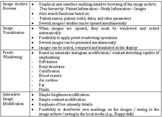
normal user. The basic operation requirements are presented in Table 1.
Table 1.
Basic image visualization requirements.
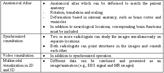
3.3. Advanced Visualization Requirements
In order to utilize more the digital nature of medical images,
advanced image processing and visualization techniques can be applied
(see Table 2). These tools can be applied in normal radiological
diagnosis by selecting appropriate preset windowing functions. In
addition, various details on the images can be efficiently emphasized
using interactive adaptive histogram equalization; original image and the
equalized image are superimposed and the transparency of the equalized
image is altered interactively. Such presentation combines the original
appearance of the image and is capable of emphasizing small low intensity
lesions and details. Another important tool is measurement; lines,
circles and polygons can be drawn on top of the images and their
dimensions can be calculated based on imaging parameters.
Table 2.
Advanced image visualization requirements.

Segmentation appears to be a key technique in order to enable
volumetric analysis, weight calculation, and 3D visualization. In
general, the purpose of segmentation is to recognize and classify
different tissues, organs and lesions on the image by using computer.
Because images consist of voxels with known dimensions, it is possible to
calculate volumes and weights of segmented structures. It is also
necessary to store segmented images to the image archive in order to
estimate e.g., disease/medication progression based on changes in volumes
or structures. Segmentation is also a key issue in electrophysiological
simulations. Appropriate anatomical models can be reconstructed based on
segmentation.
3.4. Special Requirements
Even though segmentation and volumetry are
efficient tools in estimating disease/medication progression, the total
volume of segmented lesions does not always correlate with clinical
findings. This is clearly seen in neurological diseases, such as multiple
sclerosis (MS). The poor correlation can be explained on the basis of the
locations of lesions; one small lesion can be fatal but another larger
lesion does not cause any symptoms. One small lesion located in an
important nervous pathway can create grave symptoms but at the same time
a very large lesion in not so important pathway gives rise to only
minimal symptoms. In order to improve the estimation of real lesion load,
anatomical atlases must be applied. Segmented patient images are compared
to atlas images in such way that the anatomical/functional locations of
the lesions are found out. Utilization of anatomical atlases involves
numerous problems, from which the varying anatomy of the human brain is
the most difficult one atlas images must be matched with the patient
images.
Table 3. Special
image visualization requirements.
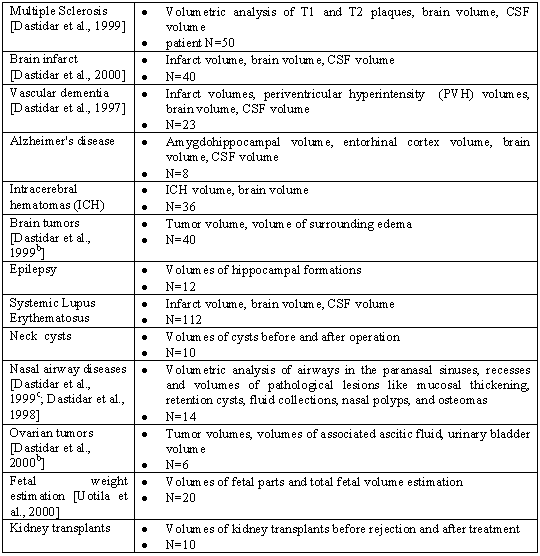
3.5. Test Studies in Clinical Environment
The developed software Anatomatic and Medimag were applied in numerous patient studies,
which are presented in Table 4. For further information for the results
on these studies, please refer to the references in the Table.
Table 4.
Test studies in clinical environment.
4. Discussion
According to our studies, 8bit gray scale hardware can be applied
efficiently in computer aided diagnosis when the entire image dynamics
are utilized in interactive windowing even though only 256 separate
intensities can be presented at a time, region of interest can appear
with better contrast when compared to conventional 12-16 bit
visualization. However, this type of interactive windowing is not yet
approved for general clinical radiology.
Automatic segmentation is not reliable due to varying anatomy hence
semiautomatic approach is recommended. However, such approach suffers
from human errors causing inter- and intraobserver
variability. In addition, some normal structures and pathological lesions
have similar kind of appearance, so it is essential to have experienced
radiologist performing the segmentation. At present, some semiautomatic
segmentation routines require fairly long time therefore the popularity
of these techniques has not yet reached daily clinical practice. In
long-term longitudinal studies as e.g., MS disease, the use of same
imaging device is recommended due to variability in resulting images. Our
experience has shown that the measured parameters at the time of imaging
and that at the time of operation differ somewhat due to existing
different circumstances on the operation table. This leads to
difficulties in calibration of the volumetric techniques.
We were able to apply the developed software in numerous different
disease studies and also in clinical practice. In addition to the
diseases mentioned earlier these software could be found useful in
various other diseases, such as gall stone diseases, tumors and
inflammatory diseases of the alimentary system, respiratory system, and
the peripheral nervous system. Also structures and tumors of the
musculoskeletal system can be quantified. Aneurysms and arteriovenous malformations can be volumetrically
estimated before and after interventional therapy/surgery.
Volumetric analysis using segmentation has become a routine procedure
in phase three drug trials in diseases like multiple sclerosis and brain
infarcts. It helps the researchers and experts to analyze their results
with more volumetric accuracy. It provides the clinicians with
measurement of the end points of drug treatment and after operations. It
also provides them with the prognosis and thus acts as a surrogate for
many clinical markers. The ease with which these
software can be used nowadays promises a future where every clinician
will have all these software at his disposal in the polyclinic room.
A decade back when computerized volumetric analysis was not yet
introduced to the routine medical analysis, manual volumetric analysis
took long tedious hours of hard work. In diseases like MS where there are
hundreds of lesions the task seemed impossible. Nowadays with the help of
these semi-automatic volumetric software, the
analysis of effects of drug and the prognosis has become much more
effective. The response to treatment in cancers like in the ovarian
tumors has become much more accurate and thus options for different
further post-operative and post-radiation treatment has become much more
effective.
With the help of the 3D software, the 3D reformations of lesions and
body structures have become more realistic and accurate. With the help of
the 3D images the effects of different lesions and tumors on the surrounding
parts can be determined in all three sagittal,
coronal and axial planes and also in different sub planes. For the
neurosurgeon the use of 3D images in navigational operations is very
helpful and has become routine in many centers.
Segmentation appears to be a key technology in computer aided
diagnosis of medical images. One of its applications is the generation of
source data for electrophysiological simulations. Voxels labeled with
tissue specific conductivities can be applied in calculating electric
field distributions. However, very accurate conductivities have not yet
been obtained, and in addition, conductivity in certain tissues is unisotropic, hence perfect models cannot be
developed.
We believe that in the near future computer-aided diagnosis will
become routine tool in radiology and other clinical fields. Before that,
some standardization is required in integrating anatomical atlases, image
analysis software, simulation tools, and PACS systems. Also teaching and
clear change in thinking attitude towards more digital diagnostic world
is required. As a conclusion for this study, the following picture
illustrates the relations of different medical research components in
order to create a complete diagnosis environment.
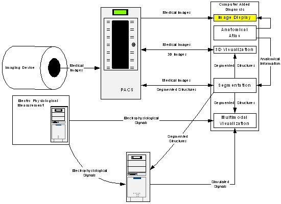 Figure 1. Image diagnosis package consists of
five main components: Image display, anatomical atlas, 3D visualization,
segmentation, and multimodal visualization. Medical images can be
uploaded from the PACS system and processed further. Segmented images can
be presented in 3D and applied in simulations together with
electrophysiological signals. In addition, original signals and
simulation results together with segmented structures can be presented as
multimodal images.
Figure 1. Image diagnosis package consists of
five main components: Image display, anatomical atlas, 3D visualization,
segmentation, and multimodal visualization. Medical images can be
uploaded from the PACS system and processed further. Segmented images can
be presented in 3D and applied in simulations together with
electrophysiological signals. In addition, original signals and
simulation results together with segmented structures can be presented as
multimodal images.
Acknowledgements
The authors wish to thank the Ragnar Granit Institute and the Ragnar
Granit Foundation. Also the Department of Radiology of the Tampere
University Hospital
is greatly acknowledged.
References
Reiner B, Siegel E, McKay P. Adoption of
alternative financing strategies to increase the diffusion of picture
archiving and communication systems into the radiology marketplace. Journal of Digital Imaging, 13(2
Suppl 1): 108-13, 2000.
Greinacher CF, Bach EF, Herforth
M, Luetke B, Seufert
G. Related Articles Computer-assisted radiology--requirements and solutions
for digital diagnostic imaging. Medical
Informatics, 15(1): 21-9, 1990.
Heinonen T, Dastidar
P, Kauppinen P, Malmivuo J, Eskola
H. Semi-automatic tool for segmentation and volumetric analysis of
medical images. Medical &
Biological Engineering & Computing, 36: 291-296, 1998.
Heinonen T, Visala
K, Blomqvist M, Eskola
H, Frey H. 3D Visualization library for multimodal medical images. Computerized Medical Imaging and
Graphics, 22(4): 267-273, 1998b.
Heinonen T, Lahtinen
A, Häkkinen V. Implementation of
Three-Dimensional EEG Brain mapping. Computers
and Biomedical Research, 32(2): 123-131, 1999.
Dastidar P, Heinonen
T, Vahvelainen T, Elovaara
I, Eskola H. Computerized volumetric analysis
of lesions in multiple sclerosis using a new semiautomatic segmentation
software. Medical & Biological
Engineering & Computing, 37: 104-107, 1999.
Dastidar P, Heinonen
T, Ahonen JP, Jehkonen
M, Molnar G. Volumetric measurements of right cerebral hemisphere
infarction: use of a semiautomatic MRI segmentation technique. Computers in Biology and Medicine.
30(1): 41-54, 2000.
Dastidar P, Kulkas
T, Heinonen T, Lahtinen
A, Ryymin P, Frey H. MR volumetry
and digital neuroanatomic mapping in vascular
dementia. Abstracts of the XVI World Congress of Neurology, Buenos
Aires, Argentina,
September 14-19, 1997.
In: Journal of the Neurological
Sciences, Supplement to vol 150. Elsevier.
S1-S367, p. S328, 1997.
Dastidar P, Heinonen
T, Virta T, Kuurne T.
Use of semiautomatic segmentation and three-dimensional reformations in
the evaluation of intracranial tumors. Medical & Biological Engineering & Computing, 37 (suppl 1): 268-269, 1999b.
Dastidar P, Numminen
J, Heinonen T, Ryymin
P, Rautiainen M, Laasonen
E. Nasal airway volumetric measurement using segmented HRCT images and
acoustic rhinometry. American Journal of Rhinology,
13(2):97-103, 1999c.
Dastidar P, Heinonen
T, Numminen J, Höckert
A, Rautiainen M. Semiautomatic segmentation of
CT images in volumetric estimation of airways in nasal cavity and paranasal sinuses. European Archives of Otorhinolaryngology,
256(4):192-198, 1998.
Dastidar P, Mäenpää
J, Heinonen T, Kuoppala
T, Van Meer M, Punnonen
R, Laasonen E. Magnetic resonance imaging based
volume estimation of ovarian tumours: use of a
segmentation and 3D reformation software. Computers in Biology and Medicine, 30: 329-340, 2000b.
Uotila J, Dastidar
P, Heinonen T, Ryymin
P, Punnonen R, Laasonen
E. Magnetic resonance imaging compared to ultrasonography
in fetal weight and volume estimation in diabetic and normal pregnancy. Acta Obstetricia
et Gynecologica Scandinavica,
vol 79: 255-259, 2000.
Laarne PH, Tenhunen-Eskelinen ML, Hyttinen JK, Eskola
HJ. Effect of EEG electrode density on dipole localization accuracy using
two realistically shaped skull resistivity
models.
Brain topography, 12(4): 249-54, 2000.
|

 Home
Current Issue
Table of Contents
Home
Current Issue
Table of Contents
