Reply to Comments Made by R. Grave
De Peralta Menendez and S. L. Gonzalez Andino
Figures
LORETA
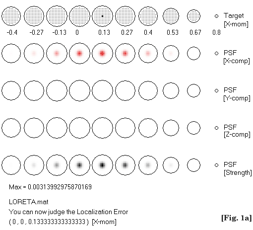
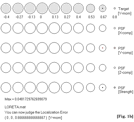
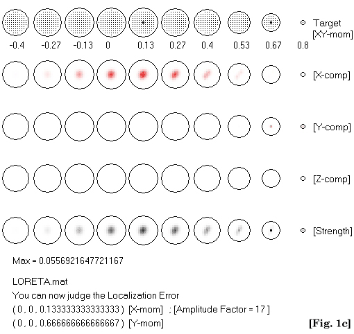
Figure 1. Rows are horizontal tomographic slices through a unit radius spherical
head. First row indicates the actual test sources and dipolar moment.
Second to fourth rows show estimated LORETA current density for each field
component (dipolar moments). Fifth row is strength of current density
field. Test sources in (1a) and (1b) have unit strength. Note that the
estimated deep source in (1a) is more blurred (about 17 times less amplitude)
than the shallow source in (1b). LORETA can resolve both simultaneously
active sources by increasing the strength of the deep source, as shown
in (1c).
Minimum Norm |
Non-regularized
 |
Regularized
 |
LORETA |
Non-regularized
 |
Regularized
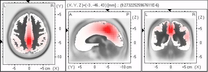 |
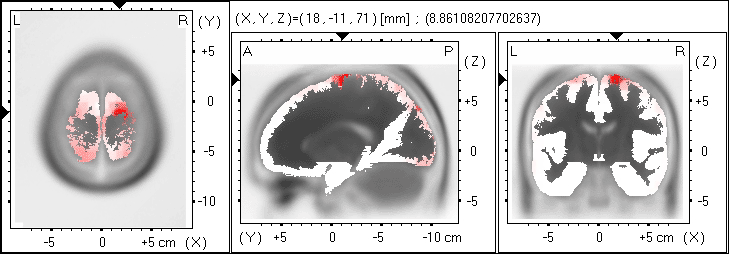
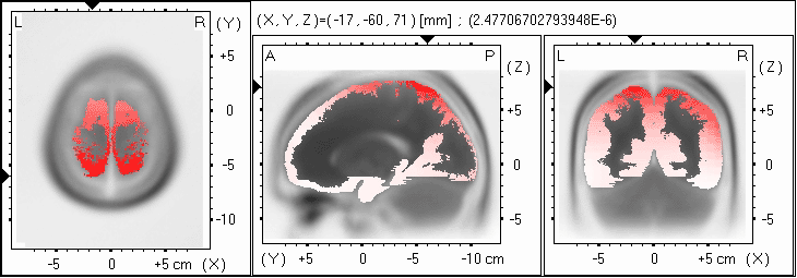
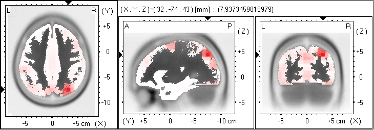
Figure 2. LORETA and minimum norm images (non-regularized and regularized) corresponding
to a test point source at Talairach coordinates (-3,-46,43). Noise was
added to the scalp voltages (ratio of variances of signal to noise (SNR)
equal to 10). Regularization was estimated via minimum cross-validation
error. Anatomy is coded in black to white. Estimated current density in
cortical grey matter is coded white (zero) to red (maximum). Location
of maximum activity is indicated numerically and by black triangles on
the coordinate axes. Estimated maximum strength is indicated numerically
(single number following Talairach coordinates). Exact localization (with
blurring) is achieved only with regularized LORETA. Other notation: (L=left;
R=right; A=anterior; P=posterior).
______________________________