|
International Journal of Bioelectromagnetism Vol. 5, No. 1, pp. 92-95, 2003. |
www.ijbem.org |
|
ST- and T-Wave in Recovery Phase Helena Hänninenab,
Jukka Nenonenbc, Markku Mäkijärviab, Toivo
Katilabc, a Division of Cardiology and bBioMag Laboratory, Helsinki University Central Hospital, Helsinki, cLaboratory of Biomedical Engineering, Helsinki University of Technology, Espoo, Finland Correspondence: H Hänninen, Helsinki University Central Hospital, Division of Cardiology, Cardiovascular Laboratory, P.O. Box 340, FIN-00029 HUCH, Finland. E-mail: helena.hanninen@hus.fi, phone +358 40 591 2261, fax +358 9 4717 4574 Abstract. ST depression is the most
common manifestation of myocardial ischemia in exercise testing. The exercise-induced
ST depression does not localize the anatomic site of coronary artery obstruction.
During the recovery phase of an exercise test the downsloping ST depression
with T-wave inversion is considered an ischemic response. Otherwise, T-wave
changes in clinical 12-lead ECG exercise testing tend to be considered nonspecific
and not utilized routinely. Body surface potential mapping studies have shown
that in addition to ST depression, changes in ST slope, T-wave amplitude,
and ST-T integral are associated with exercise-induced myocardial ischemia.
Furthermore, the ST-segment and T-wave parameters perform best in different
phases of the formal exercise test: ST segment at last workload phase and
immediately after cessation of exercise; and T-wave parameters later on during
the early recovery phase of an exercise test. This time-dependency of the
optimal performance of ST-segment and T-wave parameters should be considered.
In addition, some evidence exists that the T-wave amplitude changes in body
surface potential mapping during transient ischemia may aid in localizing
the ischemic myocardial region. The detection and localization of myocardial
ischemia could thus be enhanced by analysis of the signals over the ST segment
to the end of the T-wave and combination of the observations over different
phases of the exercise test.
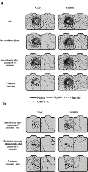
Keywords: Exercise testing; Recovery Phase; ST-T Integral; T-Wave Amplitude 1. Introduction Body surface potential mapping (BSPM), covering the whole thorax, allows comprehensive spatial analysis of body surface electrocardiogram (ECG) for detection of ischemia. The diagnostic power of 12-lead ECG in acute myocardial ischemia and myocardial infarction may not be optimal due to limited mapping area of the precordial leads. In patients with independently documented CAD, standard precordial leads do not sample the maximal ST depression in 25% [Yanowitz et al., 1982], and in 10% to 16% of patients with CAD the ST depression occurs exclusively outside the conventional precordial leads [Kubota et al. 1985]. This suggests that improved sensitivity of exercise testing may be possible with BSPM, especially in single-vessel disease. In addition to enhanced detection of ischemic ST deviation by use of BSPM, some evidence exists that body surface T-wave amplitude or combined ST-T isointegral distribution may be more sensitive in localizing the ischemia region than is ST-segment mapping alone [Ishikawa et al., 1988; Nakajima et al., 1988; Kubota et al., 1989]. In our studies in addition to ST segment we have recently focused on the algorithms based on ST-T integral area and T-wave amplitude in detection of exercise-induced myocardial ischemia. 2. ST-Segment Parameters 2.1. ST Amplitude ST-segment depression is the most common manifestation of exercise-induced myocardial ischemia, representing subendocardial ischemia. The ischemic exercise-induced ST depression does not localize the anatomic site of coronary artery obstruction neither in 12-lead ECG [Dunn et al., 1981; Mark et al., 1987] nor in BSPM [Kubota et al., 1985; Nakajima et al., 1988; Montague et al., 1990; Hänninen et al., 2001a,b]. The inability to localize exercise-induced ischemia may be due to a suggested global nature of subendocardial ischemia caused by elevated LV end-diastolic pressures [Kubota et al. 1985]. Prior MI is the most frequent cause of ST-segment elevation during exercise and is related to the presence of severe hypokinetic or akinetic left ventricular segmental wall motion [Mark et al., 1987; Yasumura et al., 1987]. In patients without a previous infarction, however, the ST-segment elevation frequently localizes the site of severe transient ischemia [Mark et al., 1987; Fletcher et al., 2001]. With progressive exercise the ischemic ST depression tends to increase [Chaitman, 1997]. In BSPM studies correlation has been high between ST changes at cessation of exercise and recovery, showing that ST integral changes reflective of myocardial ischemia persist well after exercise into the immediate recovery period [Montague et al., 1990]. Of patients with CAD, 6 % to 8 % present with ST-segment depression only during the recovery phase of an exercise test [Savage et al., 1987; Lachterman et al., 1990]. 2.2. ST Slope Morphology of ST segment, ST slope, in 12-lead ECG exercise testing currently serves as an additional criterion to ST depression ischemia detection. The same magnitude of horizontal or descending ST depression has showed higher likelihood of being ischemic than the corresponding upsloping one [Chaitman, 1997]. Horizontal ST slope < 0.7 mV/s during exercise and ST slope < 0.3 mV/s during recovery in addition to ST depression has improved the accuracy of exercise testing [Ribisl et al., 1993]. In BSPM ST slope alone, irrespective of the ST level, may perform as well as ST depression in ischemia detection during the recovery phase of an exercise test [Hänninen et al., 2001a]. Like ST depression, ST slope does not localize the ischemic myocardial region. 3. T-Wave Parameters 3.1. ST-T Integral Area and T-Wave Amplitude 12-Lead ECG Exercise Testing During the recovery phase of an exercise test downsloping ST depression with T-wave inversion is usually considered an ischemic response. Otherwise, T-wave changes in clinical 12-lead ECG exercise testing tend to be considered nonspecific and not utilized routinely [Chaitman, 1997]. Dynamics of the ST-T Integral and T-Wave Amplitude In BSPM, T-wave analysis has mainly focused on the recovery phase of an exercise test due to better signal-to-noise ratio. The dynamics of the T-wave amplitude changes during exercise testing has not been evaluated in many studies. Some evidence exists that during exercise in BSPM the T-wave amplitude changes in patients with coronary artery disease (CAD) resemble those in healthy controls, showing T-wave amplitude decrease over left upper thorax and increase over right lower thorax [Hänninen et al., in press]. The ischemic T-wave amplitude changes only become evident some minutes after the cessation of exercise, demonstrated as T-wave amplitude decrease over the left precordium in CAD patients, whereas in healthy controls corresponding changes are markedly less profound (Fig. 1). The dynamic patterns of ST-T integral area changes during the recovery resemble those of the T-wave amplitude. Dynamics of ST-T integral and T-wave amplitude changes during the immediate recovery phase of an exercise test should thus be appreciated. Localization of the Ischemic Region BSPM studies have shown that body surface T-wave potential or combined ST-T isointegral distribution may be more sensitive in localizing the ischemia region than ST-segment mapping [Ishikawa et al., 1988; Nakajima et al., 1988; Kubota et al., 1989; Hänninen et al., 2001b]. Anterior, inferior, and lateral T-wave changes have been apparent in patients with evidence of anterior, inferoposterior, or lateral ischemia, respectively, assessed by 201Tl SPECT and coronary angiography (Fig. 1) [Ishikawa et al., 1988; Nakajima et al., 1988; Kubota et al., 1990; Hänninen et al., 2001b].
Figure 1. a) From top to bottom: T-wave amplitude maps at rest, at last workload phase, immediately after cessation of stress, and at 2 minutes recovery. Left: patients with coronary artery disease (CAD), right: healthy controls (Controls). b) The phase-difference maps of the T-wave amplitude for patients with CAD and for Controls. Top: T-wave amplitude at rest was subtracted from the corresponding one immediately after cessation of stress. Middle: T-wave amplitude immediately after cessation of stress was subtracted from the one at 2 minutes recovery. Bottom: T-wave amplitude at rest was subtracted from the one at 2 minutes recovery. The step between two isocontour lines is 50 µV. 4. Discussion Body surface potential mapping as a research tool allows a comprehensive study of the electrophysiological manifestations of myocardial ischemia on body surface. The development of computer software for the analysis for body surface ECG has allowed the use of more feasible and accurate ischemia algorithms, earlier excessively time-consuming from paper ECG recordings. BSPM studies have demonstrated that in addition to ST depression, other ischemia parameters, such as ST slope, reflecting the morphology of the ST segment, and furthermore ST-T integral area and T-wave amplitude can be applied to detect exercise-induced ischemia. Neither the ST amplitude nor the ST slope can localize the culprit coronary artery during exercise. It has been suggested that this could be due to global nature of exercise-induced subendocardial ischemia. In contrast, T-wave amplitude and ST-T integral area have demonstrated ischemia localization capacity, showing ischemia-induced changes in different spatial areas of the chest surface for each patient subgroup with different ischemic regions. ST depression performs best in ischemia detection during the last workload phase and at the immediate recovery phase of an exercise test. The ischemic T-wave amplitude and ST-T integral area changes enhance during the immediate recovery phase of an exercise test. Thus, the time-dependency of the optimal performance of ST-segment and T-wave parameters should be considered. 5. Conclusions During exercise testing, the ST-segment and T-wave ischemia indexes contain information on separate physiological stages of ischemia development and resolution. The detection of myocardial ischemia could thus be enhanced by analysis of the signals over the ST segment to the end of the T-wave and combination of the observations over different phases of the exercise test. References Chaitman B. Exercise testing, in Heart disease A textbook of cardiovascular medicine. Braunwald E, Editor. Philadelphia: W.B. Saunders Company, 1997, 153-176. Dunn RF, Freedman B, Bailey IK, Uren RF, Kelly DT. Localization of coronary artery disease with exercise electrocardiography: correlation with thallium-201 myocardial perfusion scanning. American Journal of Cardiology, 48: 837-843, 1981. Fletcher GF, Balady GJ, Amsterdam EA, Chaitman B, Eckel R, Fleg J, Froelicher VF, Leon AS, Piña IL, Rodney R, Simons-Morton DA, Williams MA, Bazzarre T. Exercise standards for testing and training: a statement for healthcare professionals from the American Heart Association. Circulation, 104: 1694-1740, 2001. Hänninen H, Takala P, Mäkijärvi M, Korhonen P, Oikarinen L, Simelius K, Nenonen J, Katila T, Toivonen L. ST segment level and slope in exercise-induced myocardial ischemia evaluated with body surface potential mapping. American Journal of Cardiology, 88:1152-1156, 2001a. Hänninen H, Takala P, Mäkijärvi M, Montonen J, Korhonen P, Oikarinen L, Simelius K, Nenonen J, Katila T, Toivonen L. Recording locations in multichannel magnetocardiography and body surface potential mapping sensitive for regional exercise-induced myocardial ischemia. Basic Research in Cardiology, 96:405-414, 2001b. Hänninen H, Takala P, Rantonen J, Mäkijärvi M, Virtanen K, Nenonen J, Katila T, Toivonen L. ST-T integral and T-wave amplitude in detection of exercise-induced myocardial ischemia evaluated with body surface potential mapping. Journal of Electrocardiology, in press. Ishikawa T, Watabe S, Yamada Y, Miyachi K, Sakai Y, Ito A, Sotobata I. New diagnostic evidence on the T wave map indicating involved coronary artery in patients with angina pectoris. Circulation, 77: 301-310, 1988. Kubota I, Ikeda K, Ohyama T, Yamaki M, Kawashima S, Igarashi A, Tsuiki K, Yasui S. Body surface distributions of ST segment changes after exercise in effort angina pectoris without myocardial infarction. American Heart Journal, 110: 949-955, 1985. Kubota I, Hanashima K, Ikeda K, Tsuiki K, Yasui S. Detection of diseased coronary artery by exercise ST-T maps in patients with effort angina pectoris, single-vessel disease, and normal ST-T wave on electrocardiogram at rest. Circulation, 80: 120-127, 1989. Lachterman B, Lehmann KG, Abrahamson D, Froelicher VF. ¡±Recovery only¡± ST-segment depression and the predictive accuracy of the exercise test. Annals of Internal Medicine, 112: 11-16, 1990. Mark DB, Hlatky MA, Lee KL, Harrell FE, Califf RM, Pryor DB. Localizing coronary artery obstructions with the exercise treadmill test. Annals of Internal Medicine, 106: 53-55, 1987. Montague T, Witkowski F, Miller R, et al: Exercise body surface potential mapping in single and multiple coronary artery disease. Chest, 97: 1333-1342, 1990. Nakajima T, Kawakubo K, Toda I, Mashima S, Ohtake T, Iio M, Sugimoto T. ST-T isointegral analysis of exercise stress body surface mapping for identifying ischemic areas in patients with angina pectoris. American Heart Journal, 115: 1013-1021, 1988. Ribisl PM, Liu J, Mousa I, Herbert WG, Miranda CP, Froning JN, Froelicher VF. Comparison of computed ST criteria for diagnosis of severe coronary artery disease. American Journal of Cardiology, 71: 546-551, 1993. Savage MP, Squires LS, Hopkins JT, Raichlen JS, Park CH, Chung EK. Usefulness of ST-segment depression as a sign of coronary artery disease when confined to the postexercise recovery period. American Journal of Cardiology, 60: 1405-1406, 1987. Yanowitz F, Vincent M, Lux RL, Merchant M, Green LS, Abildskov JA. Application of body surface mapping to exercise testing: S-T80 isoarea maps in patients with coronary artery disease. American Journal of Cardiology, 50: 1109-1113, 1982. Yasumura S, Kubota I, Ikeda K, Yamaki M, Tsuiki K, Yasui S. ST segment changes in exercise body surface mapping after myocardial infarction in patients with isolated left anterior descending coronary artery disease. American Heart Journal, 114: 1120-1128, 1987.
© International Society for Bioelectromagnetism
|