|
International Journal of Bioelectromagnetism Vol. 5, No. 1, pp. 65-66, 2003. |
www.ijbem.org |
|
Influences on the Parallel Conductance Camilla
Carlssona, Lars-Åke Brodina, Göran
Källnerb, Jan Hultmanc, Håkan Elmqvista aDepartment of Medical Engineering, Karolinska
Institutet, Huddinge, Sweden Correspondence: C Carlsson,
Department of Medical Engineering,
Karolinska Institutet, Novum plan 4, S-141 86 Huddinge, Sweden.
Abstract.
The objectives were to investigate if tissues surrounding the heart
contribute to the parallel conductance in a large animal model, contradictory
to mice models, and if placement and number of electrodes changes
the recordings of the parallel conductance. Measurements were done
in five pigs during open chest surgery with the pericardium intact.
A 5-segment conductance catheter and single segment catheter were
placed inside the left ventricle to record LV volume and pressure.
Recordings were made at baseline and when the thoracic cavity surrounding
the heart was filled with physiological saline. The filling of the
thoracic cavity induced an increase of the LV volume measurements
and a decrease in measured stroke volume. The PV-loops moved significantly
to the right in the PV diagrams. The present study demonstrates that
surrounding structures contributes to the parallel conductance, at
least in the porcine model. Electrode placement, the number of electrodes
and transmitted frequency do effect the measurements of the parallel
conductance.
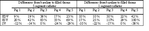
Keywords: Conductance Catheter; Parallel Conductance; Pressure-Volume Loops; LV Volume 1. Introduction The conductance catheter technique gives a continuous measurement of LV volume [Baan, 1989]. The catheter, which has several evenly spaced electrodes, is placed inside the LV. Between the most distal and proximal electrode runs a weak alternating current. The electrodes in between sense potentials from which segment conductances are derived, and the LV-volume is then derived from the sum of the segment conductances: [Baan, 1989]. A part of the current will propagate to the myocardium and surrounding tissues, even though the conductivity of blood is 3 times higher than that of the myocardium. The conductance catheter will therefore not only measure the LV blood pool but also conductance derived from the tissues surrounding the LV and changes in these may result in major measurement errors. It has been reported from mice studies that the parallel conductance derives mainly from the LV wall [Georgakopoulos and Kass, 2000]. It is desirable to know if this also applies to larger animal such as the pig. The objectives were to investigate if tissues surrounding the heart contribute to the parallel conductance in a large animal model, contradictory to mice models, and if placement and number of electrodes changes the recordings of the parallel conductance. This is especially important if you aim to use the dual-frequency method for calibration of the parallel conductance. 2. Material and Methods A single segment conductance catheter (15.5 kHz) equipped with a high bandwidth pressure sensor [Carlsson, 2001, Söderqvist, 2001] were used together with a 5-segement conductance catheter (20 kHz) (Leycom Sigma 5DF) connected to a Leycom Sigma-5 signal-conditioner processor. In vitro measurements were performed to make sure that the catheters did not interfere with each other. In vivo measurements were done in five pigs during open chest surgery with the pericardium kept intact. Both catheters were placed inside the left ventricle to record LV volume and pressure. Hemodynamic data were acquired in apnea in end-expiration to minimise the effects of intrathoracic pressure variations. After finishing the preparation procedure the animals were allowed to stabilise for approximately 20 minutes. Throughout all recordings measurements with echocardiography were made in order to estimate ejection fractions. Baseline recordings were made during stable conditions with minor parts of the anterior heart wall exposed to air. The thoracic cavity surrounding the heart was then filled with physiological saline having a higher conductivity than blood. The saline had the same temperature as the animal 's body temperature. During the filling and at baseline pressure-volume recordings were made. All signals were low pass filtered with a digital IIR 13-pole Butterworth low pass filter having a cut off frequency of 43 Hz. 3. Results The filling of the compartments surrounding the heart induced an increase of the left ventricular volume measurements and a decrease in measured stroke volume, see table 1. The PV-loops moved significantly to the right in the PV diagrams. Table 1. Changes in EDV, ESV and SV during filling of the thoracic cavity.
4. Discussion and Conclusions The reports from mice studies show that the parallel conductance derives mainly from the LV wall [Georgakopoulos and Kass, 2000]. This fact simplifies the procedure of two-frequency measurements. In contradiction to the results from mice studies our results shows that in the porcine model you cannot assume that the current will mainly stay within the LV blood pool and wall. The large differences between the five animals can be explained by the different hemodynamic conditions and also that different amounts of saline were poured into the thoracic cavities. The level of saline covering the heart was the same in all experiments. The differences between the two catheters is probably a result of different electrical fields due to different electrode spacing, placement and the number of electrodes and also the transmitted frequencies are different. We believe that the difference between our results and those from the mice studies may be attributed not only to different anatomic shapes but also to electrode placement and the relationships between the size of the animal, the electrodes and electrode spacing. The electric field might not be equal in the two settings. The present study demonstrates that surrounding structures contributes to the parallel conductance, at least in the porcine model. Electrode placement, the number of electrodes and transmitted frequency do effect the measurements of the parallel conductance. Our results are important to consider when measuring on larger animals and humans. References Baan J et al. Calibration and application of the conductance catheter for ventricular volume measurements, Automedica, vol. 11, pp. 357-365, 1989. Carlsson C et al. Initial experience with a thin single segment conductance catheter. In proceedins of the 23rd Annual International Conference of the IEEE/EMBS, 2001. Georgakopoulos D and Kass DA. Estimation of parallel conductance by dual-frequency conductance catheter in mice. Am J Physiol Heart Circ Physiol. 279(1):H443-50, Jul 2000. Söderqvist et al. Designof a single segment conductance catheter for measurements of left ventricular volume. In proceedins of the 23rd Annual International Conference of the IEEE/EMBS, 2001.
© International Society for Bioelectromagnetism
|