|
International Journal of Bioelectromagnetism Vol. 5, No. 1, p. 373, 2003. |
www.ijbem.org |
|
Effects of Different Atrial Pacing
Modes
A. Kutarski, A. Glowniak, D. Szczesniak, and P. Rucinski.
Department of Cardiology, University Medical School of Lublin, Poland Abstract. Analysis of high gain, signal-averaged (SA) ECG is an
accepted method evaluating abnormalities of atrial repolarization presence
of late potentials (ALP), predictive for atrial arrhythmias. It was proved
recently, that location of atrial lead significantly modifies atrial activation
and influences upon atrial arrhythmia recurrence.
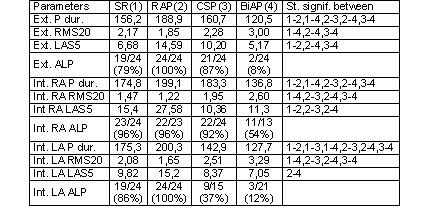
The aim of our study was to estimate the effect of different modes of atrial pacing on signal-averaged P wave recorded from external (conventional) and from intraatrial leads. Methods. Examination was performed during implantation of biatrial (BiA) pacing systems in 24 pts. External signals (SA-E) were obtained from conventional Franks orthogonal leads. Intraatrial signals were recorded separately from right (SA-RA) and left atrium (SA-LA), using bipolar pacing leads placed in RAA and CS and ventricular lead placed temporarily in LRA position. Signals were gathered during sinus rhythm (SR), right atrial appendage (RAP), coronary sinus (CSP) and biatrial (BiAP) pacing. Signals were filtered and recorded by means of Codax SAI-IK amplifier, digitized by A/D converter and stored on PC. P wave duration (Pdur), root mean square voltages of the last 20 ms of P wave (RMS20) and duration of low amplitude signal < 5mV (LAS5) were considered. Diagnostic criteria of ALP were: Pdur > 125 ms and RMS20 < 2,40 mV. Results are shown in Table 1. Conclusions. 1. RAP prolongs SA P wave duration both in external and intraatrial recordings and duration of low amplitude signal (LAS5) and distinctly decreases root mean square voltages of the last 20 ms of P wave (RMS20) in comparison to SR recorded from conventional and intraatrial leads. RAP increases the occurrence of ALP in both external and internal SAECG. 2. CS pacing favorable modifies SA time domain of left atrium. It significantly shortens P duration, distinctly increases RMS20, decreases duration of LAS5 and eliminates atrial late potentials in most of patients in comparison to SR. It does not deteriorate right atrium activation in comparison to SR. 3. BiAP shortens P duration, increases RMS20, eliminates atrial late potentials in most of patients and decreases duration of LAS5 in comparison to SR. This effect occurs in both right and left atrium and confirms beneficial effects of BiAP on atrial excitation explaining its antiarrhythmic effect. 4. Evaluation of SA intraatrial potentials yields more information about local conduction disturbances with micro-voltage oscillations during final part of atrial excitation (low RMS20 and prolonged LAS5) than conventional technique and seems to be a promising tool for evaluation of new resynchronising atrial pacing modes.
© International Society for Bioelectromagnetism
|