 |
International Journal of Bioelectromagnetism Vol. 5, No. 1, pp. 337-339, 2003. |
www.ijbem.org |
|
Body Surface Isopotential Maps to
Detect Minor István Préda, Mihály Medvegy,
Pierre Savard, and Réginald Nadeau Cardiovascular Research Group of Hungarian Academy of Sciences
and Semmelweis University, Budapest, Hungary. 1. Introduction The identification of NQMI is of paramount clinical importance, because it is more frequently associated with future UA, malignant arrhythmias, and sudden cardiac death than is QMI.1 The diagnosis of acute NQMI is generally based on clinical observations, laboratory test results, and acute ST-T changes on the ECG.2 However, ECG changes are missing when chronic NQMI is being considered. Therefore, attempts have been made to diagnose chronic NQMI through other noninvasive methods, such as echocardiography,3 thallium scintigraphy,4 radionuclide ventriculography,4 and body surface potential mapping (BSPM).5 - 9 The use of BSPM may detect electrical abnormalities such as the loss of electrical potential or altered activation sequence during depolarization. Potential loss is the reduction in the electrical activity of the heart due to a functional or structural lesion that can arise from limited necrosis or ischemia. These electrical abnormalities involving ³ 1 cardiac region correspond to myocardial damage insufficient to cause a Q wave on the standard 12-lead ECG. However, BSPM can be suitable to detect these abnormalities because of its higher spatial resolution. The position of the initial potential minimum, as suggested by Osugi et al5 and Hirai et al,6 or differences in isoarea maps, as suggested by De Ambroggi et al,7,8 were helpful in the diagnosis of old anterior and inferior NQMIs (without more accurate localization), but sensitivity was low. The aim of the present retrospective study was to analyze body surface isopotential maps in clinically proved NQMI during the chronic phase and to identify characteristic features specific to each cardiac region. 2. Methods All of the patients were hospitalized and were from the Hopital du Sacré-Coeur de Montréal. The NQMI group consisted of 45 patients (32 men and 13 women, age range 41 to 75 years, mean age 61 years), all of whom had enzymatically proved NQMI. The other reference group, the QMI group, consisted of 70 patients with remote QMI (32 anterior: 28 men and 4 women, age range 33 to 76 years, mean age 55 years; 38 inferoposterior localization: 33 men and 5 women, age range 48 to 69 years, mean age 60 years). All patients underwent coronary angiography and ventriculography, and 79 patients underwent exercise9 Tl scintigraphy. The normal control group consisted of 24 healthy adults with a normal ECG (16 men and 8 women, age range 17 to 38 years, mean age 31 years). Recording and signal processing of the body surface potentials were performed according to the Montreal system (63 unipolar leads, amplifier 0.05 to 200 Hz, signal averaging 52 seconds).10 Different types of body surface maps (isopotential, isoarea, and departure) were generated. For numerical evaluation of the features, the averaged potential value for each pathological region and the zi score for each individual NQMI patient were computed [zi=(individual potential value-normal average potential value at the same site and time)/SD of this normal average]. The specified electrode site and normalized time point of the characteristic features are given in Table 1. Individual timing was normalized [time in the individual casexaverage normal QRS duration/individual QRS duration]. The average QRS duration in our normal control group was 83±18 ms. The sensitivity, specificity, and positive and negative predictive values of the characteristic isopotential map features were evaluated and compared with different types of departure maps. Concordances were evaluated between the localized sites of potential losses through the use of isopotential maps and noninvasive or invasive cardiac methods.11 3. Results Table 1. Time, site and value of isopotential map features in different regions with potential loss, corresponding averaged normal values, difference between pathological and normal values, and sensitivity z1 score.
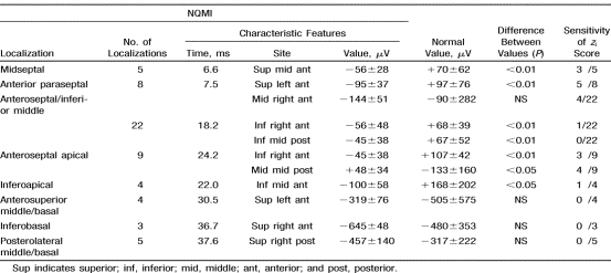
Characteristic isopotential map features were found in 40 of 44 NQMIs.
The numerical values of the pathological features with the corresponding
average normal values, the significance level of the differences,
and the sensitivity of the significant (>>1.96) zi
scores are displayed for each region in Table 1. The sensitivity,
specificity, and positive and negative predictive values of the characteristic
BSPM changes in the NQMI group were 91%, 88%, 93%, and 84%, respectively.
The pathological changes (>±2 SD) of the first third QRS and QRST
isoarea departure maps resulted in lower specificity (50% to 54%,
P<0.05. 3.1. What Does the Standard 12-Lead ECG Reveal in NQMI? According to BSPM features, the potential losses of different regions resulted in minor changes at different times during the QRS interval. The initial depolarization was involved only in cases of midseptal and anterior paraseptal potential losses: a slight r-wave reduction was revealed in leads V1 and V2. However, true pathological Q waves were seen in the leads situated above them (according to the leads V1' and V2'). For anteroseptal/inferior middle and anteroseptal apical potential losses, the r wave in leads V1 to V3 was narrower, similar to the right-sided leads. For inferoapical and inferobasal sites, a notch could appear in the inferior ECG leads. For the anterosuperior middle/basal sites, an r-wave reduction of varying degrees could be seen in leads V2 to V4. For posterolateral middle/basal sites, the r wave in leads V1 and V2 was wider and a little taller, and the real posterior leads had a wider Q wave because of the persistence of the back-sided minimum. 3.2. How Were the NQMI Features Revealed in QMI? The initial superior minimum of the midseptal and anterior paraseptal potential loss appeared in anterior QMI where the septum was involved according to the ECG (QS in leads V1 and V2, 21 of 32 cases). In extensive anterior QMI, the isopotential maps showed an extended right anterior negativity that did not allow us to discriminate among anteroseptal/inferior middle, anteroseptal apical, and anterosuperior middle/basal lesions (12 of 32 cases). The breakthrough arrived from above in 6 anterior QMI cases as in the anterosuperior middle/basal NQMI lesion. The clearly inferior QMI (22 of 38) cases showed similar inferior negativity as was seen in inferoapical NQMI. The characteristic right upper late negativity of the inferior basal NQMI location was not revealed in QMI cases. The characteristic late, back-sided minimum of posterolateral basal NQMI was even later in 10 cases with a tall R wave in lead V2. In summary, some of the pathological NQMI changes appeared in QMI, but in general, the larger extent of the necrosis masked the subtle pathological signs of the smaller regions. References 1. Goldberg RJ, Gore JM, Alpert JS, Dalen JE. Non-Q wave myocardial infarction: recent changes in occurrence and prognosis: a community-wide perspective. Am Heart J. 1987;113:273. 2. Kornreich F, Montague TJ, Rautaharju PM. Identification of first acute Q wave and non-Q-wave myocardial infarction by multivariate analysis of body surface potential maps. Circulation. 1991;84:24422453. 3. Arvan S, Varat MA. Two-dimensional echocardiography versus surface electrocardiography for the diagnosis of acute non-Q-wave myocardial infarction. Am Heart J. 1985;110:4449. 4. Wahl JM, Hakki AH, Iskandrian AS, Yacone L. Scintigraphic characterization of Q wave and non-Q-wave acute myocardial infarction. Am Heart J. 1985;109:769775. 5. Osugi J, Ohta T, Toyama J, Takatsu F, Nagaya T, Yamada K. Body surface isopotential maps in old inferior myocardial infarction undetectable by 12 lead electrocardiogram. J Electrocardiol. 1984;17:5562. 6. Hirai M, Ohta T, Kinoshita A, Toyama J, Nagaya T, Yamada K. Body surface isopotential maps in old anterior myocardial infarction undetectable by 12 lead electrocardiograms. Am Heart J. 1984;108:975982. 7. De Ambroggi L, Bertoni T, Rabbia C, Landolina M. Body surface potential maps in old inferior myocardial infarction: assessment of diagnostic criteria. J Electrocardiol. 1986;19:225234. 8. De Ambroggi L, Bertoni T, Breghi M, Marconi M, Mosca M. Diagnostic value of body surface potential mapping in old anterior non-Q myocardial infarction. J Electrocardiol. 1988;21:321329. 9. Vincent GM, Abildskov JA, Burgess MJ, Millar K, Lux RL, Wyatt RF. Diagnosis of old myocardial infarction by body surface isopotential mapping. Am J Cardiol. 1977;39:510515. 10. Savard P, Ackaoui A, Gulrajani R, Nadeau RA, Roberge FA, Guardo R, Dubé B. Localization of cardiac ectopic activity in man by a single moving dipole: comparison of different computation techniques. J Electrocardiol. 1985;18:211222. 11. Medvegy M, Préda I, Savard P, Pintér A, Tremblay G, Nasmith JB, Palisaitis D, Nadeau RA. New body surface isopotential map evaluation method to detect minor potential losses in non-Q-wave myocardial infarction. Circulation. 2000; 101:1115-1121.
© International Society for Bioelectromagnetism
|

