|
International Journal of Bioelectromagnetism Vol. 5, No. 1, pp. 304-306, 2003. |
www.ijbem.org |
|
ST Segment and T Wave Manifestations
of Wojciech
Zareba and Arthur J. Moss Heart Research Follow-up Program, Cardiology Unit, University of Rochester, Rochester, USA Correspondence: Heart Research Follow-up Program,
Cardiology Unit, University of Rochester, Box 653, 601 Elmwood Ave.
Abstract. LQTS is caused
by mutations of cardiac ion channel genes or related proteins and
ECG manifestation of the disorder is distinctive for different genetic
types of LQTS. Careful analysis of repolarization duration and morphology
in static or dynamic conditions provides vast opportunity to enhance
process of diagnosing and risk stratification of LQTS patients.
Distinct resting and dynamic ECG patterns are observed in different
genetic types of LQTS and this phenotypic presentation allows identifying
affected individuals as well as patients prone to develop arrhythmias.
1. Genetic Types of LQTS Long QT syndrome (LQTS) is a congenital disorder characterized by a prolongation of the QT interval on electrocardiogram (ECG) and a propensity to ventricular tachyarrhythmias, which may lead to syncope, cardiac arrest, or sudden death [Moss 1991; Zareba 1998]. LQTS is caused by mutations of cardiac ion channel genes or related proteins and, so far, 7 specific causative genes have been identified. Table 1 shows specific types of LQTS and respective genetic abnormalities causing them. Table 1. Genetic types of the long QT syndrome. LQTS Type Chromosomal
Gene
Ionic Channel
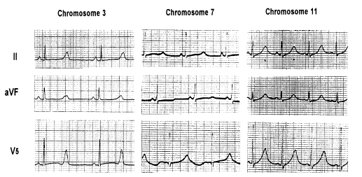
Romano-Ward Syndrome Jervell and Lang-Nielsen JLN1 11p15 KCNE1 IKsJLN2 21q22 KCNE2 IKs QT prolongation in LQTS is due to overload of myocardial cells with positively charged ions during ventricular repolarization. In LQT1, LQT2, LQT5, LQT6, and LQT7 types, potassium ion channels are blocked or open with a delay or for shorter period of time than in normally functioning channels, leading to decreased potassium outward current and prolonged repolarization. In LQT3, caused by mutations of the SCN5A sodium channel gene, persistent inward sodium current contributes to prolonged repolarization. Recently, mutation in ankyrin-B (a member of a family of versatile membrane adapters) was found to cause LQT4. Mutation of ankyrin-B results in disruption in the cellular organization of the sodium pump, the sodium/calcium exchanger, and inositol-1,4,5-trisphosphate receptors. Ankyrin-B mutation also leads to altered Ca2+ signaling in adult cardiomyocytes. It is estimated that LQT1 and LQT2 account for the majority (87%) of cases of LQTS with known genotype; LQT3 accounts for 8%, whereas LQT4, LQT5, LQT6 and LQT7 are very rare accounting for less than 5% of LQTS cases [Splawski 2000]. Homozygous KCNE1 and KCNE2 mutations are associated with congenital deafness (Jervell and Lange-Nielsen syndrome) and account for less than 1% of LQTS. There are more than 300 different mutations of these genes that have been found so far. 2. Phenotypic Presentation of LQTS by Genotype There is significant phenotypic variation of the electrocardiographic findings (T wave morphology), factors triggering cardiac events, and risk of cardiac events depending upon which gene and which mutations are involved. We have identified different ECG T-wave patterns in the LQT1, LQT2, and LQT3 forms of LQTS [Moss 1995]. Genotypic data were used to define each family member as affected or unaffected with LQTS. LQTS carriers had longer QT intervals (QTonset -c, QTpeak-c, or QTc) than unaffected family members (P<0.01). Each of the LQTS genotypes was associated with somewhat distinctive ECG repolarization features (Table 2 and Figure 1). Table 2. Quantitative Repolarization Parameters in LQT1, LQT2, and LQT3 Carriers.
Figure 1. T wave patterns in LQT1, LQT2, and LQT3 Carriers. The QT parameters (QTonsetc, QTpeakc, QTc) were most prolonged among the LQT3 carriers. T amplitude was smallest in LQT2 carriers and T wave duration was longest in LQT1 carriers. This observation set the stage for further phenotypic characterization of LQTS patients and flat, notched T wave is now recognized as a typical feature of LQT2. Remote peaked T wave with long (and sometimes somewhat elevated) ST segment is characteristic for LQT3. LQT1 carriers usually show normal looking T wave however with prolonged T wave duration almost eliminating ST segment. It is important to add that subsequent analyses demonstrated that T wave patterns vary substantially within genetic type of LQTS and also vary depending on age of studied individuals. 3. Differential Dynamics of Repolarization by Genotype QT duration and T wave morphology changes in response to variety of stimuli and conditions, which may include exercise, emotion, and action of some drugs. Differences in the sensitivity of the genotype of the congenital long QT syndrome to sympathetic stimulation (administration of epinephrine) has been studied by Shimizu and coworkers. They found that the QTc was prolonged and remained prolonged at steady state conditions of epinephrine in LQT1 patients [Noda et al. 2002]. Epinephrine also prolonged the QTc at peak of epinephrine in LQT2 patients, but this shortened to baseline levels at steady state. The QTc was much less prolonged at peak of epinephrine in LQT3 and controls than in LQT1 and LQT2 patients, and shortened to the baseline levels at steady state. This observation was further extended in a subsequent study from the same group where LQT1 patients with different penetrance of the disorder were studied [Shimizu 2003]. LQT1 carriers with borderline QTc could be identified based on response to epinephrine infusion causing substantial QTc prolongation with changes in repolarization morphology. Furthermore, symptomatic patients were presenting with more prolonged QTc in response to epinephrine than asymptomatic patients. Recently Takenaka et al. [2003] reported that QTc and Tpec were significantly prolonged during exercise in LQT1 patients (599+/-54 ms and 215+/-46 ms, respectively) with morphological change into a broad-based T-wave pattern. In contrast, exercise produced a prominent notch on the descending limb of the T wave, with no significant changes in the QTc and Tpec (502+/-82 ms and 163+/-86 ms: n=19) in LQT2 patients. Tpe interval increases during exercise in LQT1 but not in LQT2, which may partially account for the finding that fatal cardiac events in LQT1 are more often associated with exercise. Repolarization duration and morphology could also be changed differentially by beta-blocker therapy in LQT1 and LQT patients, as demonstrated in body surface mapping [Shimizu 2002]. They found that beta-blockade under normal sympathetic tone produces a greater decrease in TDR in the LQT1 form than in the LQT2 form, explaining the superior effectiveness of beta-blockers in LQT1 versus LQT2. Beta-blockers also suppress the influence of sympathetic stimulation in increasing TDR and SDR equally in LQT1 and LQT2 syndrome. 4. Summary Careful analysis of repolarization duration and morphology in static or dynamic conditions provides vast opportunity to enhance process of diagnosing and risk stratification of LQTS patients. Distinct resting and dynamic ECG patterns are observed in different genetic types of LQTS and this phenotypic presentation allows identifying affected individuals as well as patients prone to develop arrhythmias. References Moss AJ, Schwartz PJ, Crampton RS: The long QT syndrome. Prospective longitudinal study of 328 families. Circulation 1991; 84:1136-44 Moss AJ, Zareba W, Benhorin J, Locati EH, Hall WJ, Robinson JL, Schwartz, PJ, Towbin JA, Vincent GM, Lehmann MH, Keating MT, MacCluer JW, Timothy KW. Electrocardiographic T-wave patterns in genetically distinct forms of the hereditary long QT syndrome. Circulation 1995;92:2929-2934. Noda T, Takaki H, Kurita T, Suyama K, Nagaya N, Taguchi A, Aihara N, Kamakura S, Sunagawa K, Nakamura K, Ohe T, Horie M, Napolitano C, Towbin JA, Priori SG, Shimizu W. Gene-specific response of dynamic ventricular repolarization to sympathetic stimulation in LQT1, LQT2 and LQT3 forms of congenital long QT syndrome. Eur Heart J 2002;23:975-83 Shimizu W, Noda T, Takaki H, Kurita T, Nagaya N, Satomi K, Suyama K, Aihara N, Kamakura S, Sunagawa K, Echigo S, Nakamura K, Ohe T, Towbin JA, Napolitano C, Priori SG. Epinephrine unmasks latent mutation carriers with LQT1 form of congenital long-QT syndrome. J Am Coll Cardiol 2003;41:633-42. Shimizu W, Tanabe Y, Aiba T, Inagaki M, Kurita T, Suyama K, Nagaya N, Taguchi A, Aihara N, Sunagawa K, Nakamura K, Ohe T, Towbin JA, Priori SG, Kamakura S. Differential effects of beta-blockade on dispersion of repolarization in the absence and presence of sympathetic stimulation between the LQT1 and LQT2 forms of congenital long QT syndrome. J Am Coll Cardiol 2002;39:1984-91 Splawski Splawski I, Shen J, Timothy KW, Lehmann MH, Priori S, Robinson JL, Moss AJ, Schwartz PJ, Towbin JA, Vincent GM, Keating MT. Spectrum of mutations in long-QT syndrome genes. KVLQT1, HERG, SCN5A, KCNE1, and KCNE2. Circulation 2000 Sep 5;102(10):1178-85 Takenaka K, Tomohiko A, Shimizu W, Kobori A, Tomonori N, Otani H, Kubota T, Takaki H, Kamakura S, Horie M. Exercise stress test amplifies genotype-phenotype correlation in the LQT1 and LQT2 forms of the long-QT syndrome. Circulation 2003;107:838-844. Zareba W, Moss AJ, Schwartz PJ, Vincent GM, Robinson JL, Priori SG, Benhorin J, Locati EH, Towbin JA, Keating MT, Lehmann MH, Hall WJ:. Influence of the genotype on the clinical course of the long QT syndrome. N Engl J Med 1998;339:960-5.
© International Society for Bioelectromagnetism
|
|||||||||||||||||||||||||||||||||||||||||||||||||||||