|
International Journal of Bioelectromagnetism Vol. 5, No. 1, pp. 274-275, 2003. |
www.ijbem.org |
|
Action Potential
Prolongation and Contractile Dysfunction Hsin-Pao Pana, Jeng
Weic, Kuan-Yii Chenb, Tzu-Cheng Shihb, Cheng-I
Linab aInstitutes of Pharmacology & Physiology, National Defense Medical
Center, Taipei
Correspondence: CI Lin, Institute of Physiology, National
Defense Medical Center, Neihu 114, P.O. Box 90048-503, Taipei, Taiwan. Abstract. We studied the
altered action potential (AP) and contraction of left atrium isolated from
Bio 14.6 myopathic Syrian hamsters (37~52 week-old) and age-matched F1B healthy
control hamsters. Steady-state (driven at 2 Hz) twitch force and action potential
as well as post-rest potentiation of contraction (PRPC) after different rest
intervals (2~60 s) were recorded. It was found that the myopathic atria could
be divided into 2 subgroups: the dilated subgroup had significantly larger
atria size correlated with a longer APD90, while the APD90
of non-dilated subgroup was similar to that of healthy control. The PRPC vs.
rest-interval curve was significantly smaller in dilated myopathic atria than
in healthy control atria. CPA significantly inhibited the PRPC vs. rest interval
curve in healthy atria and in non-dilated myopathic atria. Our results suggest
the existence of subtypes with different characteristic alterations in action
potential and contraction in the strain Bio 14.6 myopathic hamsters.
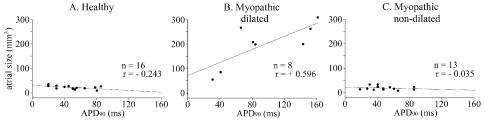
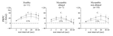
Keywords: Action Potential; Cardiomyopathic Syrian Hamster; Cyclopiazonic Acid (CPA); Left Atrial Dilatation; Post-Rest Potentiation of Contraction (PRPC); Rest Interval 1. Introduction In our previous study [Chen et al., 2001] on left atrium isolated from 2 groups of myopathic Syrian hamsters (Biobreeders Bio 14.6, 17~27 and 40~42 week-old), we found that 38% of older myopathic atria were severely dilated and action potential duration (APD) drastically prolonged as compared to younger myopathic atria and age-matched healthy controls (F1B). The aim of the present study was to extend our experiments to larger scales of myopathic hamster atria (37~39 and 48~52 week-old) to observe effects of the progress of myopathy on the abnormality in the electromechanical activity. 2. Material and Methods The animals were anesthetized with pentobarbital (50 mg/kg, i.p.) and the heart quickly removed. Left atrium was isolated and driven electrically at a basic rate of 2 Hz in 37 ℃ Tyrode solution. Steady-state twitch force and post-rest potentiation of contraction (PRPC, a measure of Ca2+ accumulation in the sarcoplasmic reticulum) [Bers et al., 1993] after different rest intervals (2~60 s) were recorded by means of a force transducer. Action potentials (APs) were recorded by means of a microelectrode technique as described recently [Chen et al., 2001]. Effects of cyclopiazonic acid, a potent inhibitor of SR Ca2+ pumping ATPase [Chiesi et al., 1994], on action potential and twitch force were tested. 3. Results It was found that at age of 37~39 weeks, left atria from 29 % of myopathic hamster were already severely dilated. At the terminal stage (48~52 weeks), incidence of atrial dilatation increased to 67 % in correlation with APD prolongation, but 3 out of 9 older myopathic atria remained non-dilated with unchanged APD. When data from 21 myopathic left atria (37~52 week-old) were pooled together, myopathic atria had significantly longer APD90 (r = 0.572, P<0.05) than that of 16 healthy control atria (r = -0.243, P>0.05) (Figure 1 A). The myopathic atria could be divided into 2 subgroups: the dilated subgroup had significantly larger atrial size correlated with a longer APD90 (n=8, r = 0.596) (Figure 1B) while the APD90 of non-dilated subgroup was similar to that of healthy control (n=13, r = -0.035) (Figure 1 C). PRPCs were determined in 11 healthy and 17 myopathic atria. The descending phase of PRPC vs. rest-interval curve (20~60 s) was significantly smaller in dilated myopathic atria (n=7) than in healthy control atria (Figure 2). When changes in PRPC were expressed as % of maximum △F, CPA (10 μM) inhibited significantly the ascending phase (up to 20 s) of the PRPC vs. rest-interval curve in healthy atria and in non-dilated myopathic atria (n=10). CPA significantly inhibited PRPC only after the shortest rest interval (2 s) in dilated myopathic atria. 4. Discussion The present results in hamster left atria suggest the existence of subtypes (dilated and non-dilated) with different characteristic electromechanical defects in the strain Bio 14.6 myopathic hamsters. The results with CPA indicate that the SR function in the atrium is already impaired in younger myopathic hamsters, and more so in older myopathic hamsters. These results were in agreement with results of our recent study on ventricular papillary muscle of myopathic hamster and human ventricular myocardium [Wei et al., 2003]. Further studies on cardiomyocytes isolated from myopathic vs. healthy atria by means of whole-cell voltage-clamp technique are in progress. Acknowledgements The present study was supported by grants NSC91-2320-B016-061 and NSC90-2314-B350-001. Figure 1. Relation of atrial size (the ordinate) to action potential duration at 90% repolarization (APD90, the abscissa) in left atria obtained from healthy (n=16) and myopathic hamsters (n=21, 37~52 week-old). Myopathic atria were divided further into 2 subgroups: dilated (middle panel, n=8) and non-dilated (right panel, n=13) according to size of the left atrium. The correlation coefficient (r) for each group is indicated in each panel. Figure 2. Post-rest potentiation of contraction (PRPC) of healthy atria (left panel, n=11), dilated (middle panel, n=7) and non-dilated myopathic atria (right panel, n=10). The ordinate gives PRPC in values (mean±SE) of twitch force above the steady-state values (△ force) before (solid circles, C.) and after CPA exposure (empty circles). The abscissa gives rest intervals (from 2 to 60 s). #P<0.05, significantly different from values in healthy control. Horizontal broken lines indicate the zero PRPC. References Bers DM, Bassani RA, Bassani JW, Baudet S, Hryshko LV. Paradoxical twitch potentiation after rest in cardiac muscle: increased fractional release of SR calcium. Journal of Molecular and Cellular Cardiology. 25: 1047-1057, 1993. Chen KY, Liu HW, Lin CL, Wei J, Lin CI. Role of sarcoplasmic reticulum in the abnormal electromechanical activity of myopathic hamster atrium. In: ELECTROCARDIOLOGY 2001. Paster CA, Editor. Proceedings of the 28th International Congress of Cardiology. Atheneu, Sao Paulo, Brazil, 2001, 519~525. Chiesi M, Wrzosek A, Gryueninger S. The role of the sacoplasmic reticulum in various types of cardiomyocytes. Molecular and Cellular Biochemistry. 130: 157-171, 1994. Wei J, Liu HC, Lee FY, Lee MS, Huang CY, Pan HP, Lin CI. Role of the sarcoplasmic reticulum in altered action potential and contraction of myopathic human and hamster ventricle. Clinical and Experimental Pharmacology and Physiology. 30 (4, in press). 2003.
© International Society for Bioelectromagnetism
|