 |
International Journal of Bioelectromagnetism Vol. 5, No. 1, pp. 258-259, 2003. |
www.ijbem.org |
|
Effect of Ischemia Size on Body Surface Maps by Realistic Volume Conductor Model Tuukka Arola, Jari Hyttinen,
Jari Viik, Noriyuki Takano, and Jaakko Malmivuo Ragnar Granit Institute, Tampere University of Technology, Tampere, Finland Correspondence: TA Arola, E-mail: tuukka.arola@tut.fi Abstract. A bioelectric field simulation
software for realistic finite difference method (FDM) volume conductor models
was produced. The software utilises lead field theory and the sources are
presented as multiple dipoles. The software outputs body surface potential
maps (BSPMs). The program was utilized to test the effect of the size of subendocardial
ischemia with a realistic male torso model. The results match well to clinical
measurements and similar potential distribution patterns can be observed.
Keywords: BSPM; Forwad Problem; FDM; Ischemia; Dipole 1. Introduction A simulation software to utilize precalcualted lead fields from finite difference method (FDM) models was produced to simulate cardiac activity with dipoles in a realistically shaped volume conductor. The software is capable of representing arbitrary sized FDM data and visualizing it from three planes. It also provides functionality to insert multiple dipoles to the model manually or through automatic dipole distribution. The software calculates resulting body surface potential maps (BSPMs) by utilizing lead field theory. Ischemia can be simulated by approximating it as a double layer that can be represented as multiple dipoles. The software was utilized to simulate the effect of a subendocardial ischemia size to BSPMs. Regardless of the location of the ischemia, BSPMs are found to be quite similar in clinical results [Hänninen, 2002] and to have similar potential distribution on the body surface. The purpose of this study was to examine the effect of ischemia size to body surface potential strengths and distribution. 2. Material and Methods The volume conductor model used was a realistically shaped
inhomogeneous FDM model of a human torso that was originally created from
a stack of MR images of a male patient. The resolution of the model was The software is based on lead field theory that was introduced by Mcfee and Johnston [McFee et al, 1953]. In lead field approach, each body surface point has a lead field calculated that describes the sensitivity of the lead to the sources in the volume conductor. The lead field can be obtained by applying reciprocity theorem which states that in finite volume conductor models the detector and the source can be exchanged without detecting any change in measured amplitudes. A lead vector contributes to a surface node through equation:
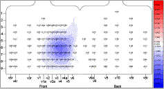
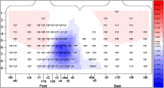
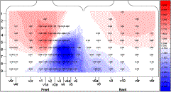
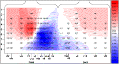
The model in this study had 117 surface points with 11732 source nodes in the heart area that had a lead vector calculated. The source model used was dipole. A dipole can represent a uniform double layer of various sizes and a larger uniform double layer can be divided into smaller sections and represented as multiple dipoles. In this study all dipoles were equal in strength. Dipoles were used to approximate a double layer representing ischemic area in myocardium. The normal activity of the heart itself is not present and only the contribution of the source dipoles representing the ischemic border area was simulated. The simulations were performed with multiple dipoles, the number ranging from 147 to 768 depending on the size of the ischemia. The ischemia was located to origin from the inferior apex of the left chamber and it spreads out to the right chamber region as the size gets larger. 3. Results Figure 1 shows the simulated BSPMs. The size was increased in non-constant steps, which is due to the FDM model and software restrictions. Figure 1 shows that the effect of ischemia causes rise in potentials on upper parts of the torso. In (a) the ischemia is represented by 147 dipoles and covering approximately 7% of the whole heart area. In (b) the number of dipoles is increased to 320 as the size is increased to 15% of the whole heart area. In (c) the area has spread to be large enough to cover parts of the right chamber surface and it is represented in total of 528 dipoles while the ischemia covers approximately 30% of the of the whole heart area. In (d) the size of the ischemia is almost half of the heart and is approximated with 768 dipoles. Positive potentials appear to the maps as the area gets large enough, in this simulation approx. 15% of the whole heart area.
Figure 1. BSPMs as the ischemia size is increased. The maps are scaled and thus comparable. Each of the potential steps represents a value range of 10% of the maximum value. (a) is the initial state where the ischemia is approximately 7% of the whole heart. In (b) and (c) the size of the ischemia was approximately 15% and 30% of the whole heart. (d) The ischemia covers almost half of the heart. 4. Discussion The results from the simulations show the same phenomenon observed in clinical measurements as the ischemia location is changed the potential distribution remains spatially constant regardless of the size of the ischemia and match well for example to those presented by Hänninen [Hänninen, 2002]. The BSPMs in Figure 1 include a small error which was due to the roughness and low resolution of the used thorax model. The artifact is seen as slight increase in upper torso potentials. The same artifact is present in all of the maps but is not visible due to the scaling and contour step size. The artifact could be reduced by using a more detailed model or by approximating the double layer with fever, evenly distributed dipoles. However, at the present state, the software is not capable of producing such layers. The software is not specialized to only this kind of simulation where the effect of the size of a double layer is studied. The software can be loaded with data of any kind and size of FDM model thus not limiting the usage to torso models only. Each of the dipoles can be individually tuned as well as automatic distribution can be used to generate approximations of double layers. There are various other features including automated scaling of the results, zooming to the model and data import and export. Other studies performed with this model and software include a study where the number of dipoles representing a double layer was studied, and a simulation of subendocardial ischemia that develops and penetrates the myocardium and turns to be transmural ischemia. References McFee R, Johnston FD. Electrocardiographic leads I. Introduction. Circulation 8:(10) 554-68. 1953.
© International Society for Bioelectromagnetism
|
 (d)
(d)
