O. Ziermann*, D. Meyer-Ebrecht*
Introduction
Many heart diseases alter form and dynamics
of the surface of the endocardium. Reconstructing the endocardium is therefore
an important task of medical image processing. Ultrasound imaging is a
low-invasive imaging modality widely available due to its low cost. Technical
advantages are the high temporal and spatial resolution. A modality for
acquisition of volumetric data sets is

Figure 1. Imaging cone in transesophageal ultrasound
fourdimesional transesophageal ultrasound. This modality uses a transducer
probe that has to be swallowed by the patient to image the heart from the
esophagus. At the tip of the probe a transducer array is positioned that
is capable of acquiring two-dimensional sequences. Rotating the tip of
the probe the imaging plane is rotated so that a cone-shaped volume can
be acquired (Fig.1). For each imaging plane an image sequence of one heart
beat is acquired. Resorting the images one obtains for each moment of the
heartbeat one volumetric dataset.
The acquisition of the whole four-dimensional dataset takes several
minutes. One problem of the imaging modality are motion artifacts that
occur due to unintentional movements of the patient during that long examination
procedure. They lead to displacements and rotations of the imaging plane
relative to their ideal positions.
Methods
Active surfaces
The analysis of endocard motion is based on the
reconstruction of a parametrized endocardial surface X(u,v) for
each moment of the heartbeat. Cohen et. al. [1] and McInerney et.al. [3]
formulated surface reconstruction as an energy-minimizing problem. An energy
function
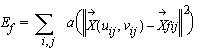
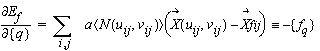
Etot=Ei+Ef for a
surface is set up consisting of an internal energy Ei
enforcing smoothness and a feature energy Ef enforcing
similarity between the surface points and corresponding feature points
computed from the image. The surface to be reconstructed relates to a local
minimum of the total energy.
 |
(1) |
 |
(2) |
X(uij,vij) are points of the surface,
Xfij
are the corresponding feature points each numbered by
i and
j.
In order to solve the problem numerically the surface has to be approximated
by a superposition of a finite number of functions. In the case of a finite
element approximation these are the piecewise polynomial formfunctions
[2]. The expansion coefficients are called knotvariables. So the approximated
surface can be written as the product of the rowvector of formfunctions
and a columnvector of knotvariables.
 |
(3) |
We follow the notation of [2] where columnvectors are symbolized with
pointed brackets, rowvectors are symbolized with braces and matrices are
symbolized with square brackets. The finite-element approximation casts
the function of the internal energy into a quadratic function of the knotvariables:
 |
(4) |
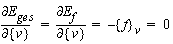
At a local minimum of the total energy the partial derivatives after
the knotvariables vanish:
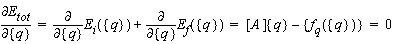 |
(5) |
where
 |
(6) |
is the vector of so called generalized feature forces. The generalized
feature forces depend on the position of the surface given by the knotvariables
through eq.3. Eq.5 is a precondition for a local minimum of the total energy.
A way of finding the local minimum of the total energy is introducing an
iteration time and descending the gradient of the total energy according
to:
 |
(7) |
This evolution equation converges to a minimum of the total energy.
The gradient descent algorithm needs to be given an initial condition to
start from. The evolution equation is solved by descretizing eq.7 with
finite differences in the time domain and integrating numerically:
 |
(8) |
The iteration can be stopped if the change between consecutive time
steps falls below a given threshold. The solution of eq.8 corresponds to
the evolution of a damped elastic membrane under the influence of a space
dependent image force computed from the image according to eq.6.
An integrated pseudomechanical model
The reconstruction of the endocardial surface
requires the correction of the above mentioned motion artifacts. Correcting
the motion artifacts on the basis of a correlation of neighbouring images
is problematic, because speckle noise is uncorrelated between neighbouring
images.
Correction of the motion artifacts and surface reconstruction are closely
related problems. On the one hand correction of the motion parameters influences
the surface to be reconstructed. On the other hand a reconstructed surface
can serve as a reference for computing the displacement of the imaging
planes [4], [5].
The problem to be solved can be put in the following words:
Compute
- a surface and
- a set of displacement parameters
so that:
- the surface is possibly smooth and
- the surface is possibly similar to the feature points.
These demands shall be put into an energy-minimising framework.
First the displacement parameters of the imaging planes must be specified.
Each imaging plane has six degrees of freedom of a solid body. Dri
and Dzi are translation coordinates
of image plane i in a cylindrical coordinate system. Dti
is a translation perpendicular to the image plane. Dai,
Dbi
and Dji are rotations around
the r-, z- respectively the t-axis. These parameters
are combined to a parameter-vector  for
each imaging plane. Feature points {rij,zij}
are computed in each imaging plane, i being an index for the imaging
plane. The index j indicates the feature points computed in the
imaging plane. The location of the feature points in space depends on the
location of the feature points in the imaging plane and the displacement
parameters of the respective imaging plane:
for
each imaging plane. Feature points {rij,zij}
are computed in each imaging plane, i being an index for the imaging
plane. The index j indicates the feature points computed in the
imaging plane. The location of the feature points in space depends on the
location of the feature points in the imaging plane and the displacement
parameters of the respective imaging plane:
 |
(9) |
where
 ,
,
 and
and
 are
rotation matrices. The requirement of similarity between the reconstructed
surface and feature points in space can be formalized in a feature energy:
are
rotation matrices. The requirement of similarity between the reconstructed
surface and feature points in space can be formalized in a feature energy:
 |
(10) |
that is depending on both, the degrees of freedom {q} of the
surface and the degrees of freedom {v} of the displacement parameters
of all imaging planes. The task of computing a surface and a set of displacement
parameters so that the surface is possibly smooth and possibly near to
the feature points can be formulated as an energy minimization problem
in the following way: Find a local minimum of a total energy consisting
of an internal energy depending on the surface and a feature energy depending
on both the surface and the displacement parameters:
 |
(11) |
The internal energy is the internal energy of an active surface. At
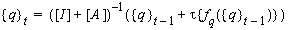
a local minimum of the total energy the derivatives after the degrees of
freedom of the surface {q} and the derivatives after the displacement
parameters {v}must vanish. The first leads to a condition of balance
similar to (eq.5). The letter leads a set of equations:
 |
(12) |
If the rotation axis of the ventricle and the rotation axis of the imaging
cone are approximately identical, the derivatives after ai,
bi
and ti are
negligible so that one can confine oneself to the computation of displacements
Dri
and Dzi
and rotation Dji
i.e. motion in the imaging plane. Eq.12 is a condition of balance of the
sum of the feature forces of one imaging plane that retroact on the imaging
plane. The term containing the derivatives after Dji
corresponds to the torque of the feature forces. The local minimum of the
total energy is again found descending the gradient of the total energy
according to:
 |
(13) |
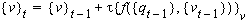 |
(14) |
Eq.13 and Eq.14 are coupled because feature forces {fq}
and {fv} depend on both the position of the imaging plane
and an the displacement parameters. This energy minimising model can be
interpreted as a pseudomechanical model of an elastic membrane coupled
to freely movable imaging planes by feature forces computed in the images
(Fig.2). In this picture the gradient descent algorithm is a damped movement
of the coupled system to its equilibrium position.

Figure 2. Pseudomechanical model
Results
The Algorithm was applied to artificial data sets
with given displacement parameters and to real data sets. Applying the
algorithm to artificial data sets both the surface and the displacement
parameters were determined correctly. In both cases displacement parameters
were determined during the first steps of the iteration. Iteration was
stopped when the difference of the surface position was below a threshold
of 1 pixel. Convergence was reached after approximately 40 iteration steps.
Fig.3 is linked with a sequence showing the evolution of the intersecting
contour during the iteration. The rigid body movement of the intersecting
contour relates to a change of the displacement parameters of the slice
plane during the iteration. Fig. 4. is linked with a sequence showing the
evolution of the surface during the iteration.

Figure 3. Temporal evolution of the intersecting contour

Figure 4. Temporal evolution of the surface
Conclusions
This paper presents an energy-minimizing algorithm
that integrates the tasks of surface detection and correction of motion
artifacts in fourdimensional transesophageal ultrasound sequences. The
problem arises whenever the raw data for a surface reconstruction scheme
is acquired tomographically. The regularization capabilities of the algorithm
can be improved if additional temporal smoothness constraints are taken
into account. Another way of enhancing stability is starting from the assumption
that unintentional movements occur on a slower time scale than heart motion
itself. One displacement vector for one heart beat i.e. one displacement
parameter per slice plane would then be sufficient. The reduced number
of displacement parameters would contribute to additional stability of
the algorithm.
References
[1] Cohen, I., Cohen, L. and Ayache, N., "Using deformable
surfaces to segment 3-d images and infer differential structures", Second
European Conference on Computer Vision, pp. 648-652, 1992.
[2] Dhatt, G. and Touzot, G., "The finite element
method displayed", John Wiley and Sons, New-York, ISBN 0-471-90110-5, 1984.
[3] McInerney, T. and Terzpopoulos, D., "A dynamic
finite-element surface model for segmentation and tracking in multidimensional
medical images with application to cardiac 4D image analysis", Computerized
Medical Imaging and Graphics, vol. 19, no. 1, p. 69-83, 1995.
[4] Schreckenberg, M., Dunkhase, K.-M., Mumm,
B. and Waldinger, J., "Verfahren zur Bewegungskompensation bei Ultraschallaufnahmen
eines Objekts", German patent filing, DE 199 03 332 .3, 1999.
[5] Ziermann, O. and Meyer-Ebrecht, D., "Integration
von Oberflächenrekonstruktion und Verschiebungskorrektur in der tomographischen
4D-Echokardiographie", Bildverarbeitung für die Medizin 2000: Algorithmen
- Systeme - Anwendungen, pp. 138-142, 2000.

 Home
Current Issue
Table of Contents
Home
Current Issue
Table of Contents
