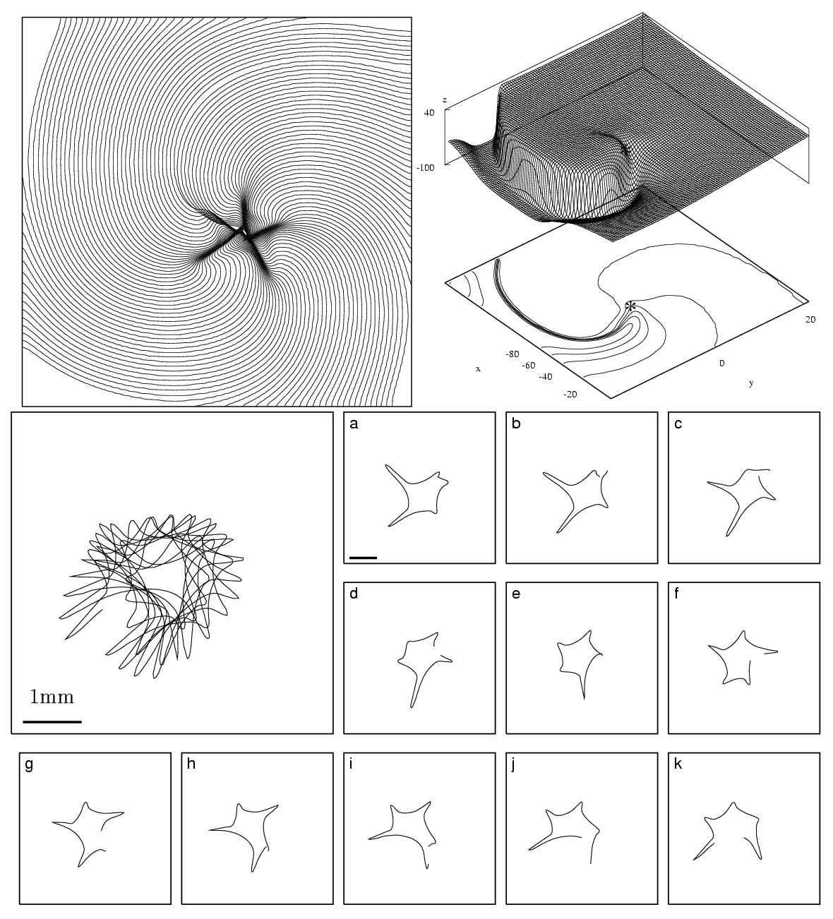
 Figure
1:
Spiral-wave solution and tip-trajectory evolution for the "normal" guinea-pig ventricular
model. (a) Isochrones of the wavefront shown every 1ms. A total of 105 isochrones are plotted
(average rotation period is approximately 100ms throughout the simulation). (b) Spatial voltage
distribution (V®
z-axis) snapshot.
The numbers at the bottom of the plot indicate the V-level
contours and the asterisk the position of the spiral tip. (c) Tip trajectory evolution for the "normal"
guinea-pig ventricular model (standard parameter values used see [5]). The region shown here is
5.5×
5.5 mm2
. The enlarged panel shows tip trajectory evolution for 1100ms starting at
t=1900ms
to remove transients. Panels a-k
show the same evolution in segments of 100ms. The horizontal
solid bar in the enlarged and the first panel is 1mm wide.
Figure
1:
Spiral-wave solution and tip-trajectory evolution for the "normal" guinea-pig ventricular
model. (a) Isochrones of the wavefront shown every 1ms. A total of 105 isochrones are plotted
(average rotation period is approximately 100ms throughout the simulation). (b) Spatial voltage
distribution (V®
z-axis) snapshot.
The numbers at the bottom of the plot indicate the V-level
contours and the asterisk the position of the spiral tip. (c) Tip trajectory evolution for the "normal"
guinea-pig ventricular model (standard parameter values used see [5]). The region shown here is
5.5×
5.5 mm2
. The enlarged panel shows tip trajectory evolution for 1100ms starting at
t=1900ms
to remove transients. Panels a-k
show the same evolution in segments of 100ms. The horizontal
solid bar in the enlarged and the first panel is 1mm wide.